Figure 1. Histological evaluation of cytokine-induced ocular inflammation in the anterior and posterior chambers, respectively. A, D: Sham injected. B, E: Interleukin (IL)-8. C, F: Tumor necrosis factor alpha (TNF-α) 24 h post-injection. C = cornea; AC = anterior chamber; V = vitreous. Magnification
= 10X. Scale bar = 100 μm.
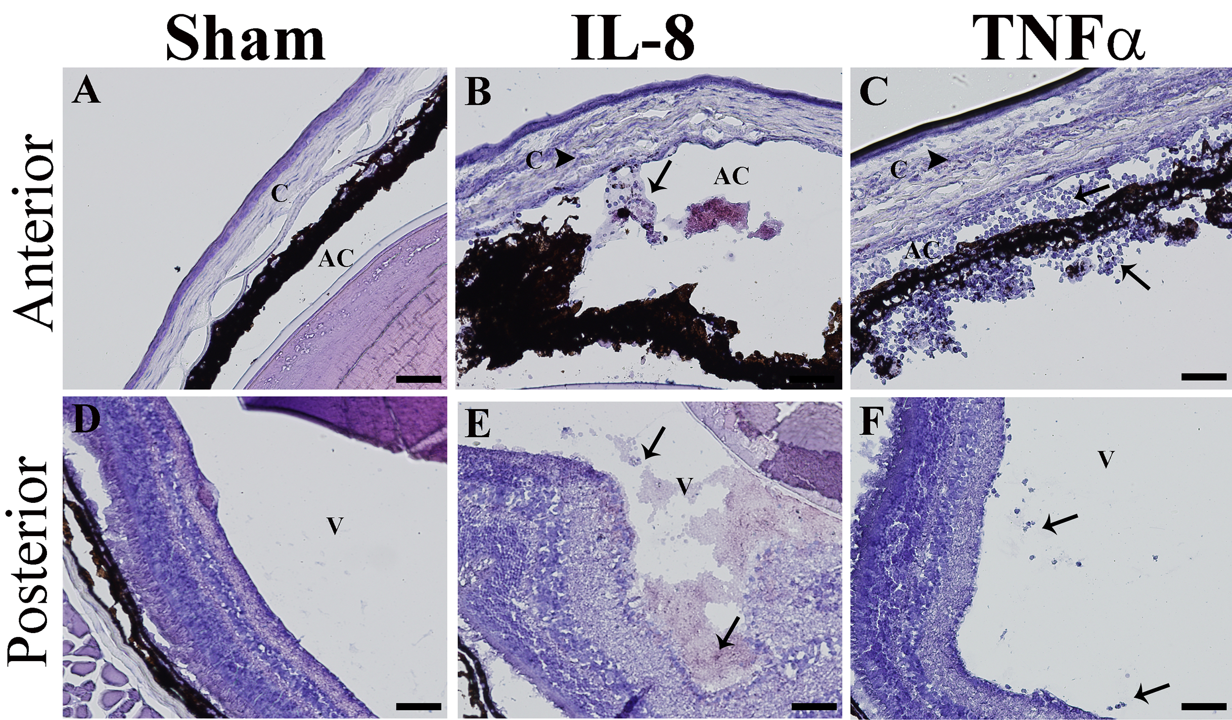
 Figure 1 of
Barliya, Mol Vis 2017; 23:922-932.
Figure 1 of
Barliya, Mol Vis 2017; 23:922-932.