Figure 5. rd10 retinas from dark-reared mice undergo slower degeneration compared with retinas from cyclic light-reared mice. A: At P42 and P67 raised in the dark, a significant number of photoreceptor cell nuclei (stained with 4',6-diamidino-2-phenylindole
(DAPI); blue) are still present (five to six rows), based on the thickness of the outer nuclear layer (ONL). Mice placed in
normal cyclic light at P42 for 4 days (P42 + 4L) undergo rapid degeneration, such that the ONL is approximately one to two
rows of nuclei at the end of day 4. pCREB (green) is detected in many, but not all, of the Müller glia (MG) in the P42 + 4L
mice. White arrows, the MG nuclei do not display pCREB staining. B: Quantification of the images from the rd10 mice raised in the dark and then exposed to light (P42 + 4L) demonstrates a general reduction of pCREB in the MG compared
with the retinas from the P42 and P67 mice maintained in the dark. Anti-SOX9 conjugated to CF555 (see Methods), final antibody
concentration, 1:2,000; anti-pCREB conjugated to Alexa Fluor 488 (anti-pCREB-Alexa Fluor 488 conjugate); final antibody concentration;
1:100. A single z slice is presented for each image. Each image represents a single mouse retina. Error bars represent the
standard error of the mean (SEM). Scale bar = 50 μm. **p = 0 .0012; ***p = 0.0002. ONL, outer nuclear layer; INL, inner nuclear
layer; GCL, ganglion cell layer.
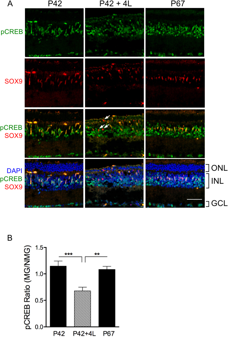
 Figure 5 of
Dong, Mol Vis 2017; 23:90-102.
Figure 5 of
Dong, Mol Vis 2017; 23:90-102.