Figure 4. Müller glia and microglia in wild-type and rd10 retinas. A: In wild-type mice, the Müller glia (MG) stained for SOX9 (red) are aligned in the INL. A microglial cell stained with Iba1
(green; marked by white arrow) is shown in the OPL; B: In the rd10 mice, the MG have moved to the inner edge of the ONL by P25. By P34, there are areas where the MG can be observed in a linear
array in the ONL, which may be preliminary to the formation of a glial seal (see Discussion). The white arrows indicate microglia.
Anti-SOX9 was conjugated to CF555 (see Methods); the final antibody concentration was 1:2,000. The anti-Iba1 was conjugated
to CF488A (see Methods); the final antibody concentration was 1:500. A single z slice is presented for each image from a single
mouse retina. INL, inner nuclear layer; ONL, outer nuclear layer, GCL, ganglion cell layer. Scale bar = 25 μm.
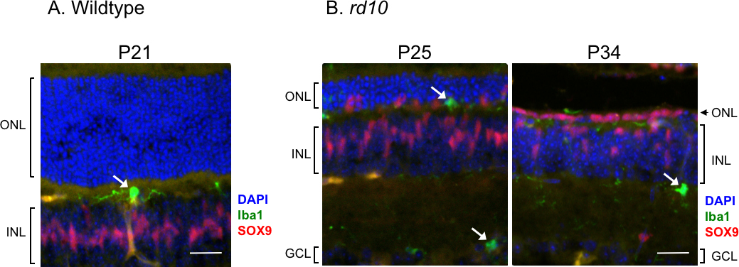
 Figure 4 of
Dong, Mol Vis 2017; 23:90-102.
Figure 4 of
Dong, Mol Vis 2017; 23:90-102.