Figure 6. Microscopic analysis of VAP-1 in human retinas. A: Immunofluorescent staining of VAP-1 in representative samples from non-diabetic control (C) and diabetic (D) donors. After fixation, retinas were stained with a specific goat anti-VAP-1 antibody (red). Nuclei were labeled with DAPI
(blue). RPE = retinal pigment epithelium; ONL = outer nuclear layer; INL = inner nuclear layer; RGL = ganglion cell layer.
B: VAP-1 colocalized with collagen IV in human diabetic retinas. a: VAP-1 immunofluorescence (red). b: collagen IV immunofluorescence
(green). c: VAP-1 (red), collagen IV (green), and nuclei (DAPI, blue). Orange fluorescence (white arrows) shows the colocalization
of both forms of fluorescence in retinal vessels (V).
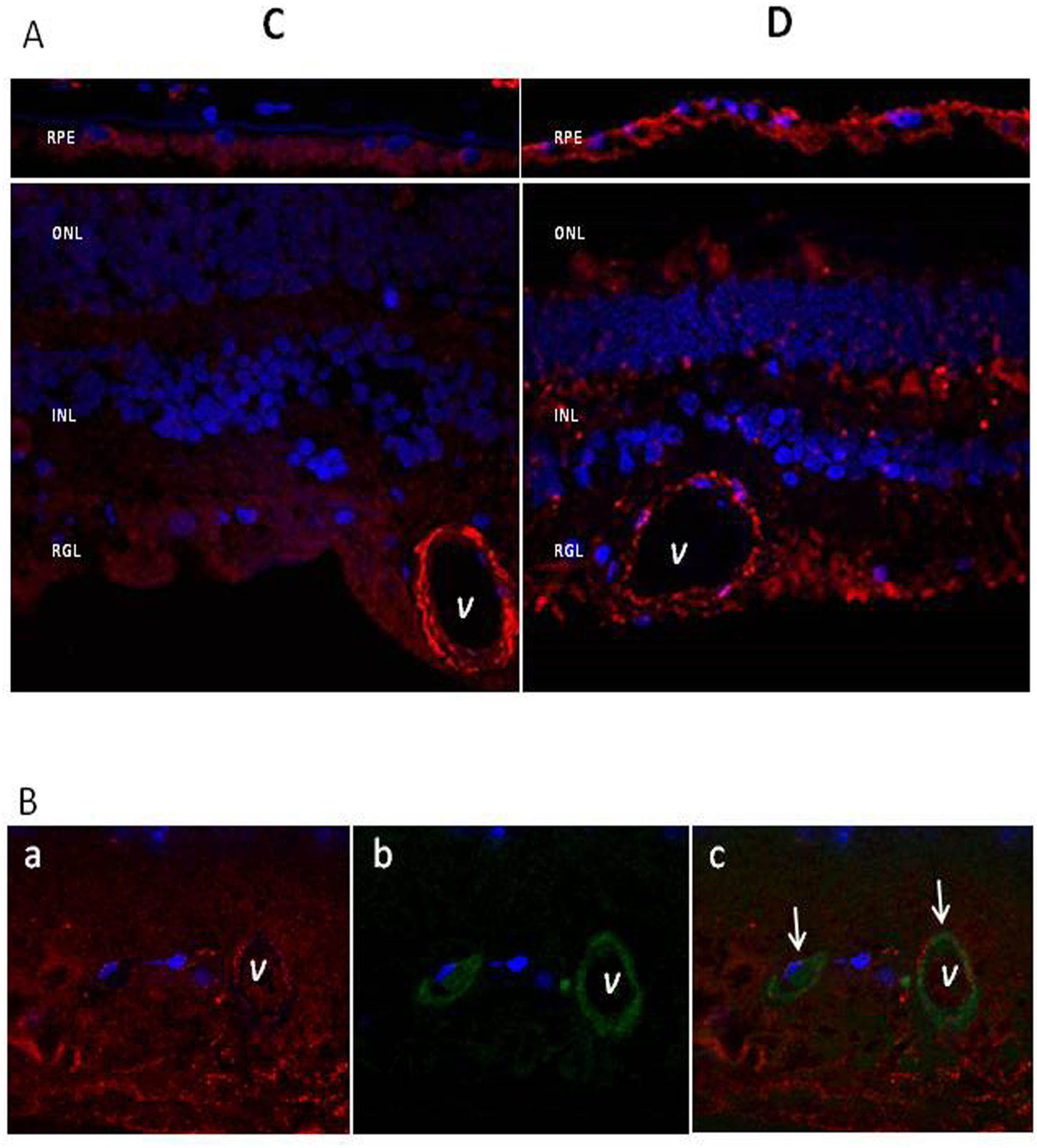
 Figure 6 of
Abu El-Asrar, Mol Vis 2017; 23:853-871.
Figure 6 of
Abu El-Asrar, Mol Vis 2017; 23:853-871.