Figure 4. PDR epiretinal membrane immunostaining. Immunohistochemical staining of VAP-1 showing immunoreactivity in vascular endothelial
cells (arrows) and stromal cells; low power (A: original magnification 25X) and high power (B: and C: original magnification 40X); stromal spindle-shaped cells expressed VAP-1 (arrows; C); double immunohistochemistry of CD45 (brown) and VAP-1 (red) showing stromal cells co-expressing CD45 and VAP-1 (arrows;
D and E; original magnification X40); immunohistochemical staining of 8-OHdG showing nuclear and cytoplasmic immunoreactivities in
vascular endothelial cells (arrows) and stromal cells (arrowheads; F: original magnification 25X).
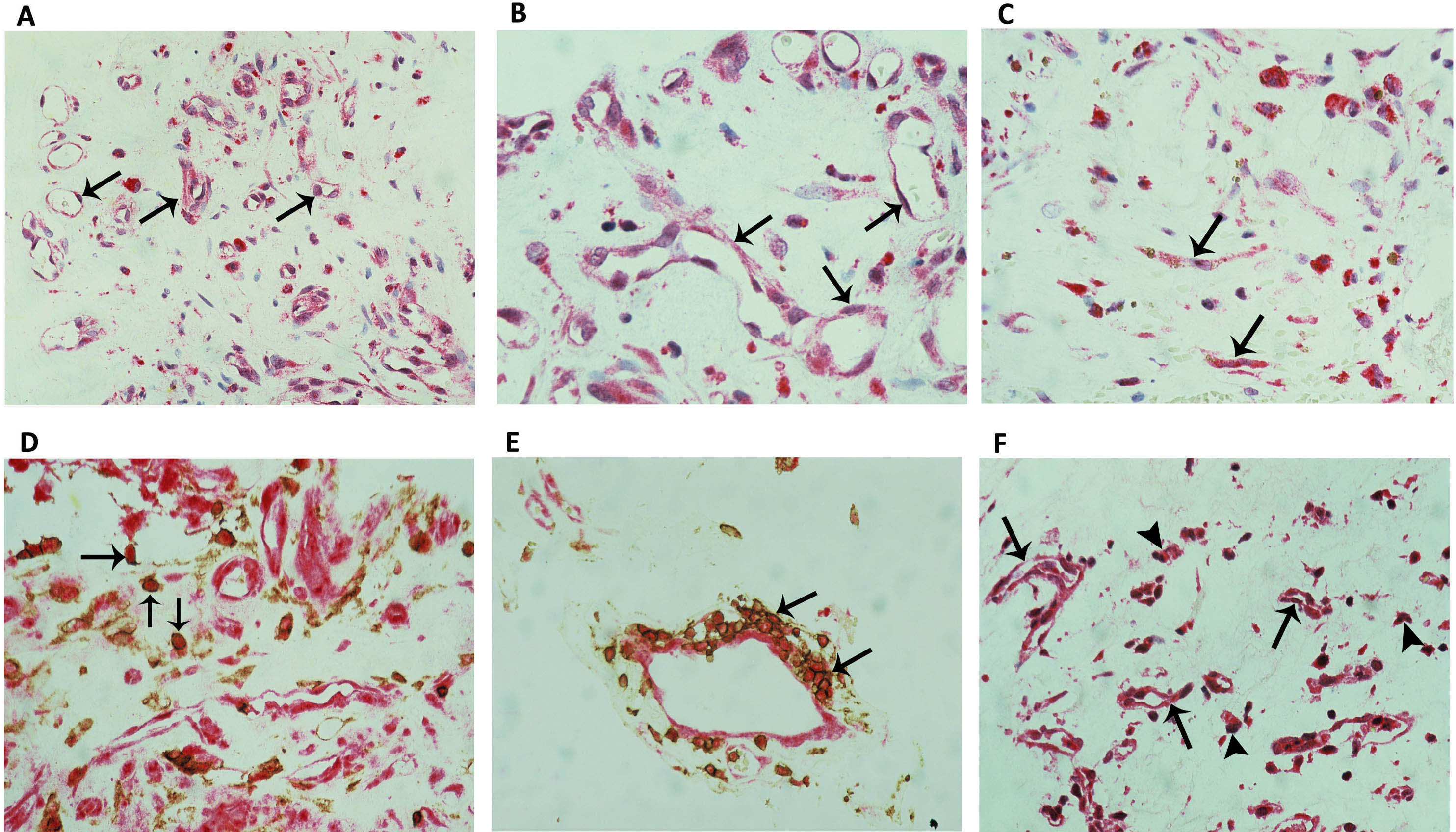
 Figure 4 of
Abu El-Asrar, Mol Vis 2017; 23:853-871.
Figure 4 of
Abu El-Asrar, Mol Vis 2017; 23:853-871.