Figure 3. PDR epiretinal membrane immunostaining. Negative control slide showing no labeling A: immunohistochemical staining of CD31 showing blood vessels positive for this endothelial cell marker (arrows; B; original magnification 25X); immunohistochemical staining of CD45 showing stromal cells positive for CD45 (arrows; C); immunohistochemical staining of α-smooth muscle actin showing immunoreactivity in myofibroblasts (arrows; D; original magnification 40X).
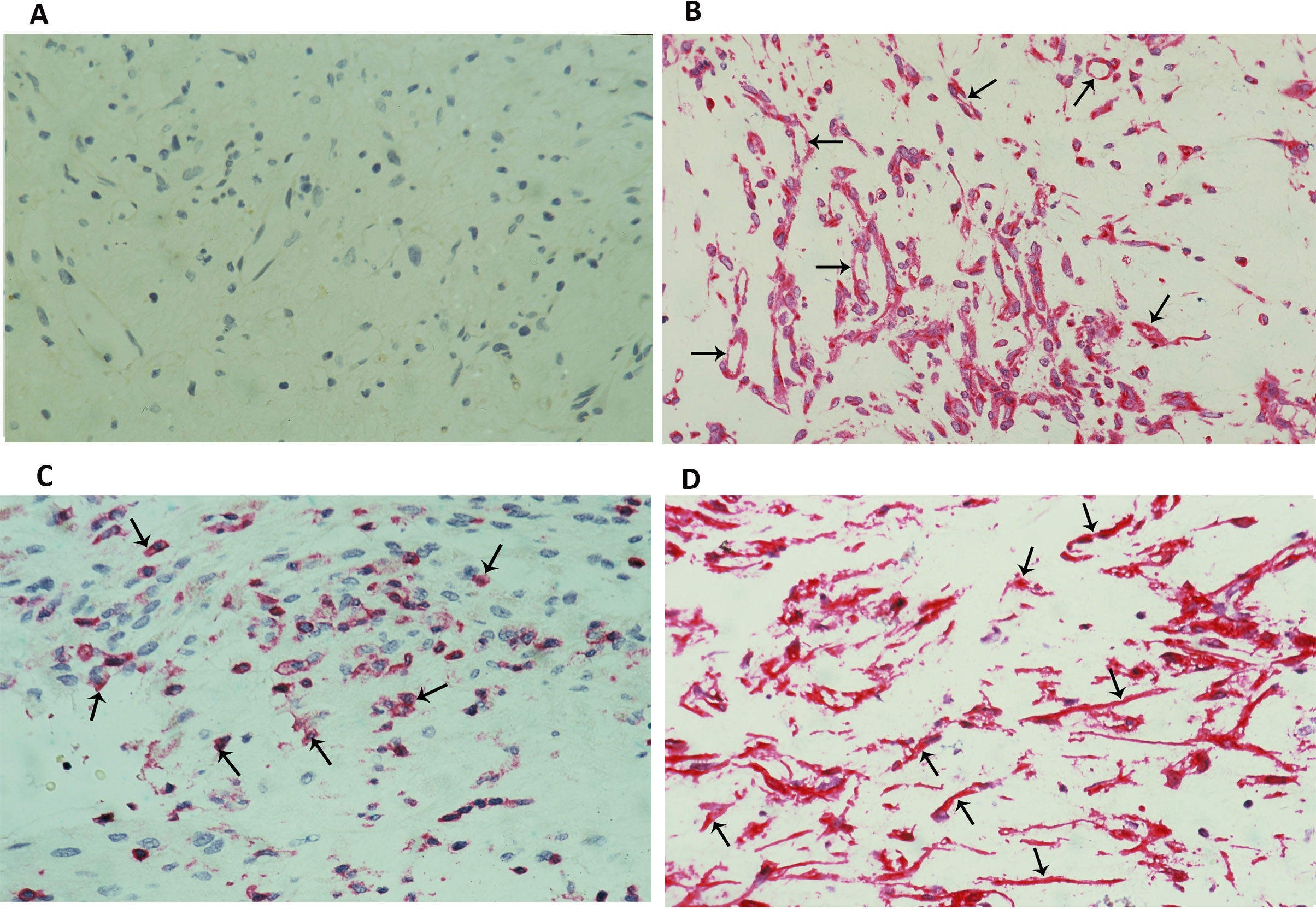
 Figure 3 of
Abu El-Asrar, Mol Vis 2017; 23:853-871.
Figure 3 of
Abu El-Asrar, Mol Vis 2017; 23:853-871.