Figure 2. Fundus images and localization of subretinal injections. A–C: The fundus images of the untreated and pilot- and subretinal PBS injection (sPBS)-treated eyes were normal. D–E: After subretinal injection of pFmCD44 green fluorescent protein (GFP) lentivirus, the fundus image was normal (D), and GFP expression was localized in the central retina (E). F: No green fluorescence was detected in the sPBS-treated eyes.
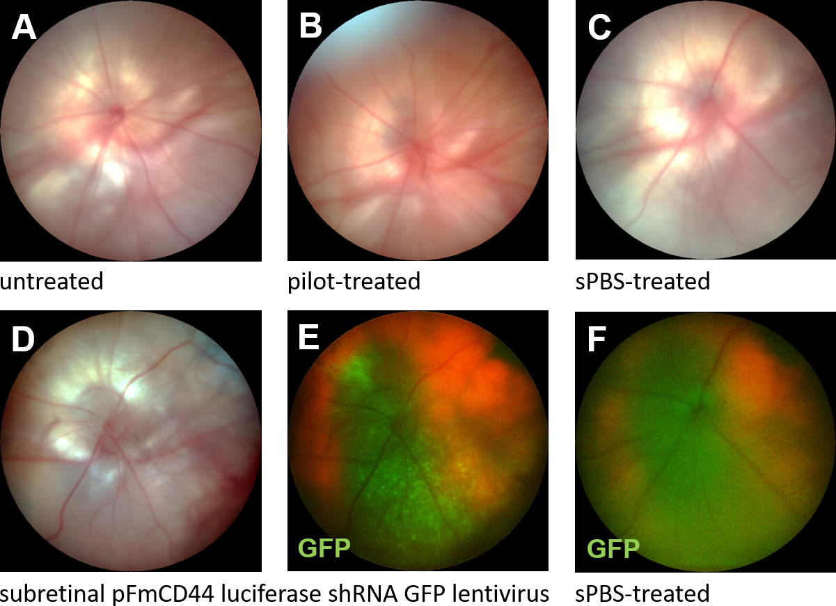
 Figure 2 of
Becker, Mol Vis 2017; 23:832-843.
Figure 2 of
Becker, Mol Vis 2017; 23:832-843.