Figure 4. Intravitreal injection of dasatinib does not induce ocular toxicity in mice. Mice received a single intravitreal injection
of dasatinib (1 µg in 1 µl dimethyl sulfoxide [DMSO]) or DMSO (1 µl; contralateral control) on P12 (oxygen-induced retinopathy,
OIR) or immediately after laser injury (choroidal neovascularization, CNV). On P17 (OIR) or 2 weeks after laser injury (CNV),
the eyes were enucleated for histological and immunohistochemical analyses. The retinal toxicity of dasatinib in mice with
CNV was assessed in the region without CNV lesions. In addition, normal mice received the same intravitreal administration
of dasatinib or DMSO. Two weeks later, the eyes were enucleated for the analysis of retinal toxicity. A: Representative images of hematoxylin and eosin (H&E)–stained retinas and quantification of the indicated ratio of A (the
distance from the ganglion cell layer to the outer edge of the inner nuclear layer) to B (the distance from the ganglion cell
layer to the outer edge of the outer nuclear layer). Data are presented as the mean ± standard error of the mean (SEM; ns=not
significant, n=4 mice per group). B: Terminal deoxynucleotidyl transferase dUTP nick end labeling (TUNEL) assay and (C) immunofluorescence staining with anti-glial fibrillary acidic protein (GFAP) immunoglobulin G (IgG) were performed on tissue
sections prepared from the eyecups of mice with OIR or CNV. In (B), DNase I-treated sections were included as positive controls. In (C), sections stained with nonspecific IgGs were included as negative controls (NC). The nuclei were stained with 4’,6-diamidino-2-phenylindole
(DAPI; blue). Representative images in (B) and (C) were selected from four independent experiments with similar results. All scale bars=50 µm.
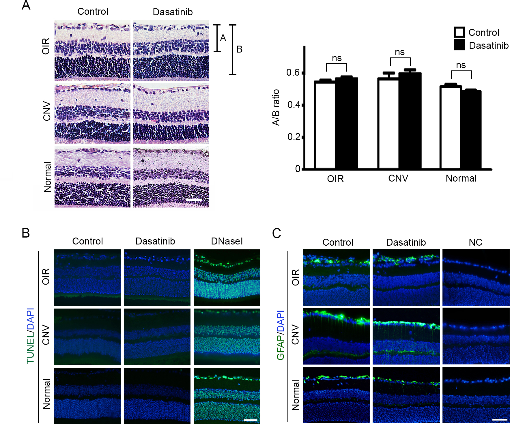
 Figure 4 of
Seo, Mol Vis 2017; 23:823-831.
Figure 4 of
Seo, Mol Vis 2017; 23:823-831.