Figure 3. Dasatinib suppresses the development of CNV in a laser-induced mouse model. A: Schematic diagram of the laser-induced choroidal neovascularization (CNV) experiment. Immediately after laser photocoagulation,
the mice received a single intravitreal injection of dasatinib (1 µg in 1 µl dimethyl sulfoxide [DMSO]) or DMSO (1 µl; contralateral
control). Two weeks later, the mice were perfused with fluorescein isothiocyanate (FITC)-labeled dextran (green), and their
posterior eyecups comprising the RPE, choroid, and sclera were stained with isolectin B4 (red), an endothelial cell marker.
B: Representative images of CNV lesions and quantification of the area and volume of the CNV lesions in the mice treated with
DMSO (control) or dasatinib. The CNV area was analyzed with measurement of the fluorescence intensity of the images with the
isolectin B4–positive area. The CNV volume was calculated as the sum of the CNV areas in confocal Z-stack images. All data
are presented as the mean ± standard error of the mean (SEM; *p<0.05, **p<0.01, n=10 mice per group). Scale bar=100 μm.
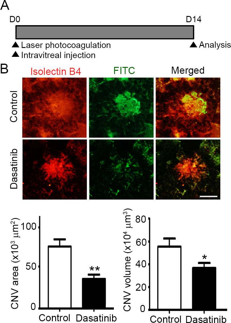
 Figure 3 of
Seo, Mol Vis 2017; 23:823-831.
Figure 3 of
Seo, Mol Vis 2017; 23:823-831.