Figure 7 of
Man, Mol Vis 2017; 23:810-822.
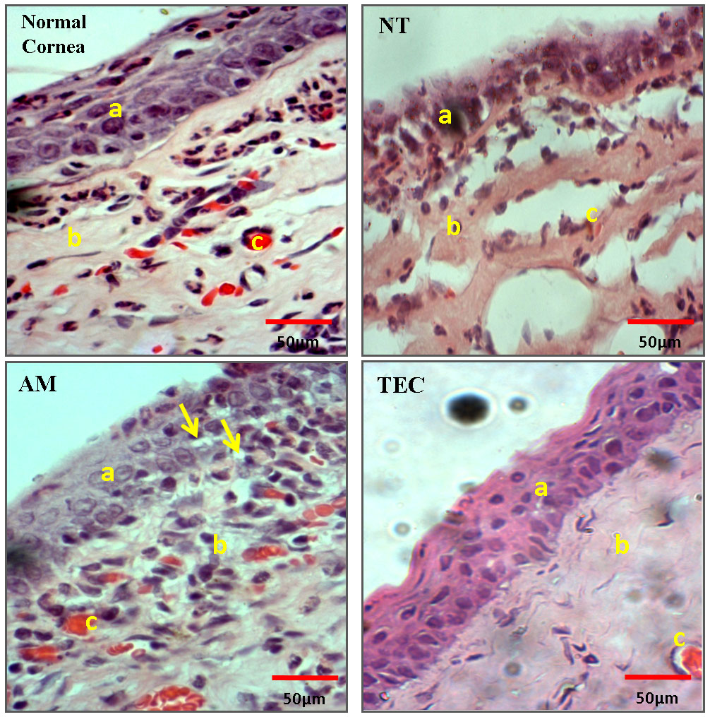
Figure 7.
Histological analysis using hematoxylin and eosin-stained normal corneal layer, NT, AM, and TEC. a: epithelial layer. b: stromal layer. c: blood vessel. Yellow arrow: goblet cells (magnification: 400x).
 Figure 7 of
Man, Mol Vis 2017; 23:810-822. Figure 7 of
Man, Mol Vis 2017; 23:810-822.
Figure 7 of
Man, Mol Vis 2017; 23:810-822. Figure 7 of
Man, Mol Vis 2017; 23:810-822.