Figure 4. Slit lamp examination of nude rat eyes showing the total epithelial defect of the corneal surface on Day 1 after corneal injury.
A representative photo of corneal defect pre-implantation showed positive fluorescein staining (green). The absence of the
green color in the rest of the photos shows that full coverage of the corneal epithelium has been achieved by Week 2. Microscopic
evaluation is also shown at 2 weeks, 4 weeks, and 8 weeks after implantation.
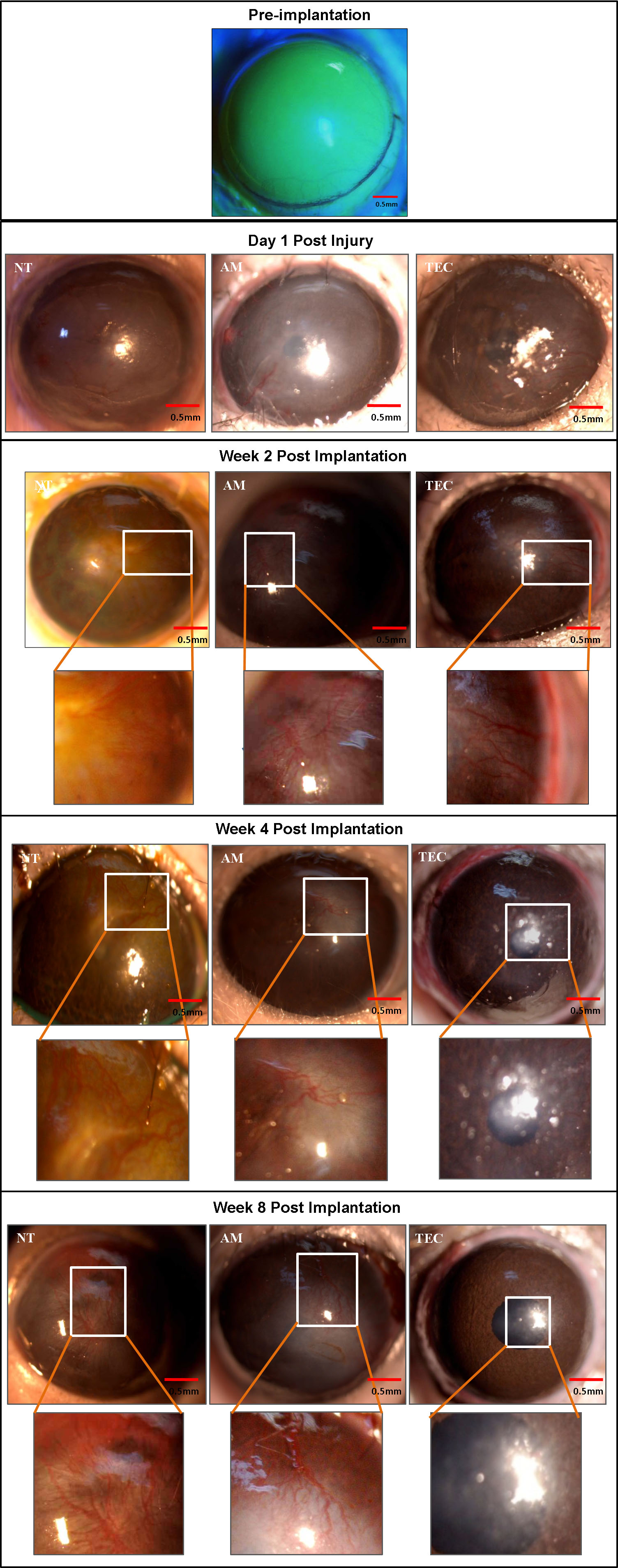
 Figure 4 of
Man, Mol Vis 2017; 23:810-822.
Figure 4 of
Man, Mol Vis 2017; 23:810-822.