Figure 2 of
Man, Mol Vis 2017; 23:810-822.
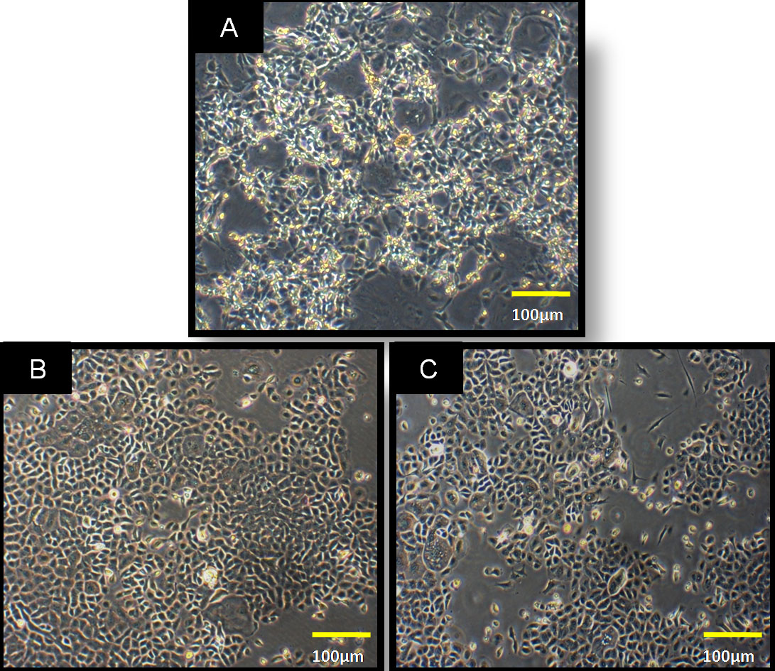
Figure 2.
Morphology of the cells.
A
: Corneal epithelial cells from the native human corneal ring samples.
B
: Uninduced BMuc after 10 d of culture.
C
: Induced BMuc cells after 10 d of induction (magnification: 40x).
 Figure 2 of
Man, Mol Vis 2017; 23:810-822.
Figure 2 of
Man, Mol Vis 2017; 23:810-822.