Figure 7. The internal structure of BM-MSC and RPE cell phagocytic activity on transmission electron microscopy. (Row 1) Bone marrow
mesenchymal stem cells (BM-MSCs) phagocytizing the photoreceptor outer segment (POS) at 24 h at different magnifications.
(Row 2) BM-MSCs phagocytizing POS for 48 h. The layer-like substance was smaller than at 24 h. (Row 3) RPE cells phagocytizing
the POS for 48 h. The layer-like substance was less and more loose at 48 h than at 24 h.
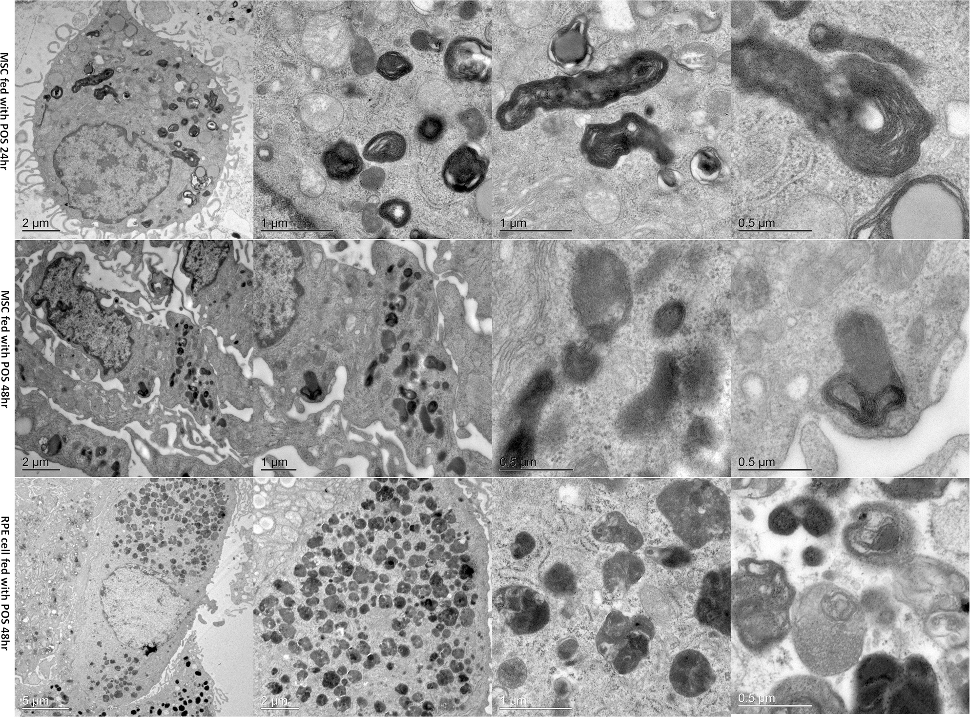
 Figure 7 of
Peng, Mol Vis 2017; 23:8-19.
Figure 7 of
Peng, Mol Vis 2017; 23:8-19.