Figure 6. The exterior structure of BM-MSC and RPE cell phagocytic activity on scanning electron microscopy. (Top left) Bone marrow
mesenchymal stem cells (BM-MSCs) were incubated with the photoreceptor outer segment (POS) for 3 h. (Top right) BM-MSCs were
incubated with the POS for 24 h. (Bottom left) RPE cells were incubated with the POS for 3 h. (Bottom right) RPE cells were
incubated with the POS for 24 h. The empty white arrows indicate the POS that bound to the cell surface. The filled white
arrows indicate microvilli. The common point of the MSC and RPE cells is the cytomembrane that protrudes around the bound
POS.
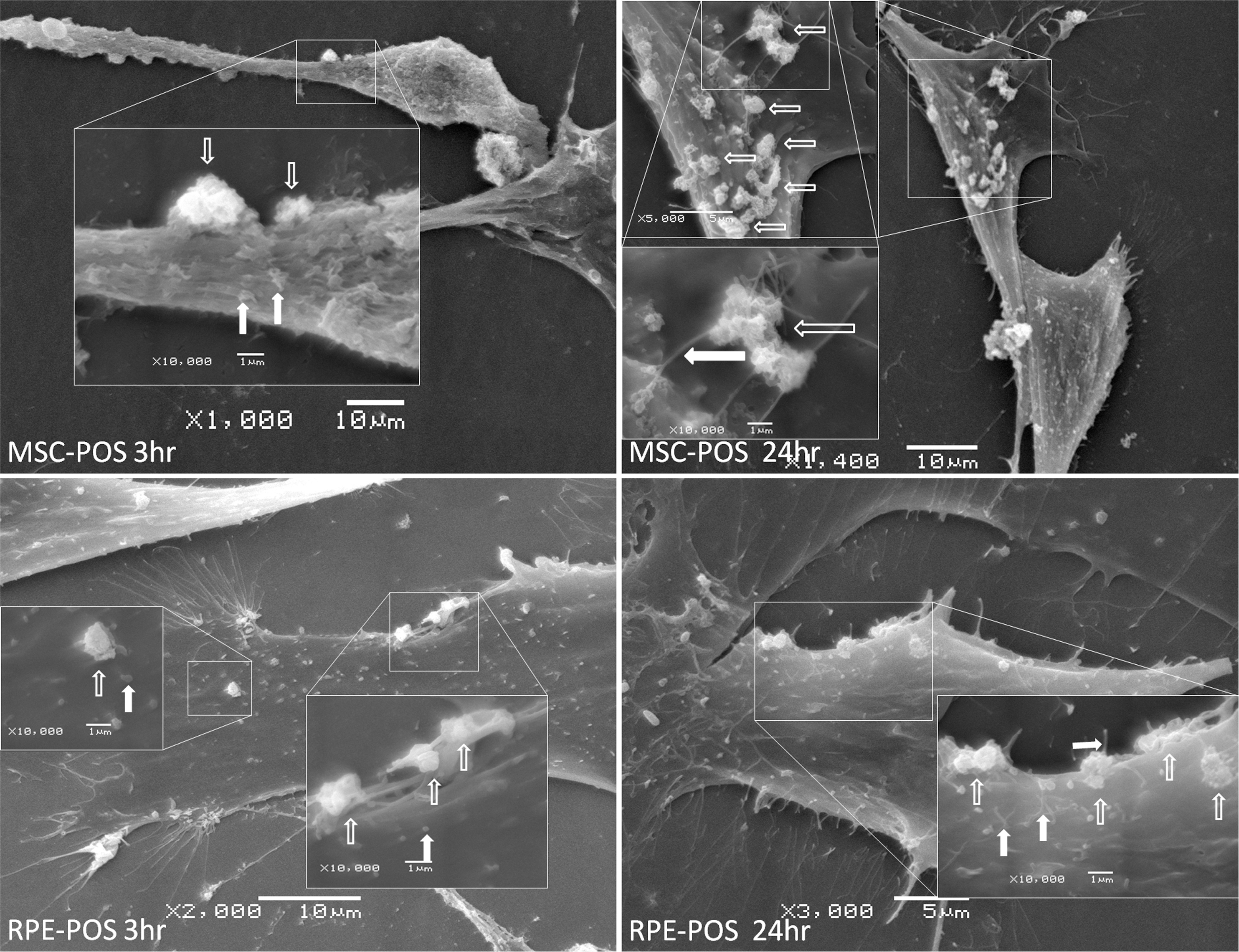
 Figure 6 of
Peng, Mol Vis 2017; 23:8-19.
Figure 6 of
Peng, Mol Vis 2017; 23:8-19.