Figure 5. Quantity of phagocytic latex beads (LBs) and photoreceptor outer segment (POS) by bone marrow mesenchymal stem cells (BM-MSCs)
and RPE cells incubated with LB and POS for different time periods. n.s. means no statistical significance between the BM-MSCs
and the RPE cells. The sampling size for each group is 3.The error bars are standard deviation (SD). The data were analyzed
with one-way ANOVA. The quantity of phagocytized latex beads did not differ between the BM-MSCs and the RPE cells from 3 to
18 h (p>0.05) but differed at other time points (p<0.05). The quantity of phagocytized POS differed between the BM-MSCs and
the RPE cells except at 3 h (p = 0.49). Before 3 h, phagocytic ability was stronger for the RPE cells than for the BM-MSCs.
After that, the situation was the reverse.
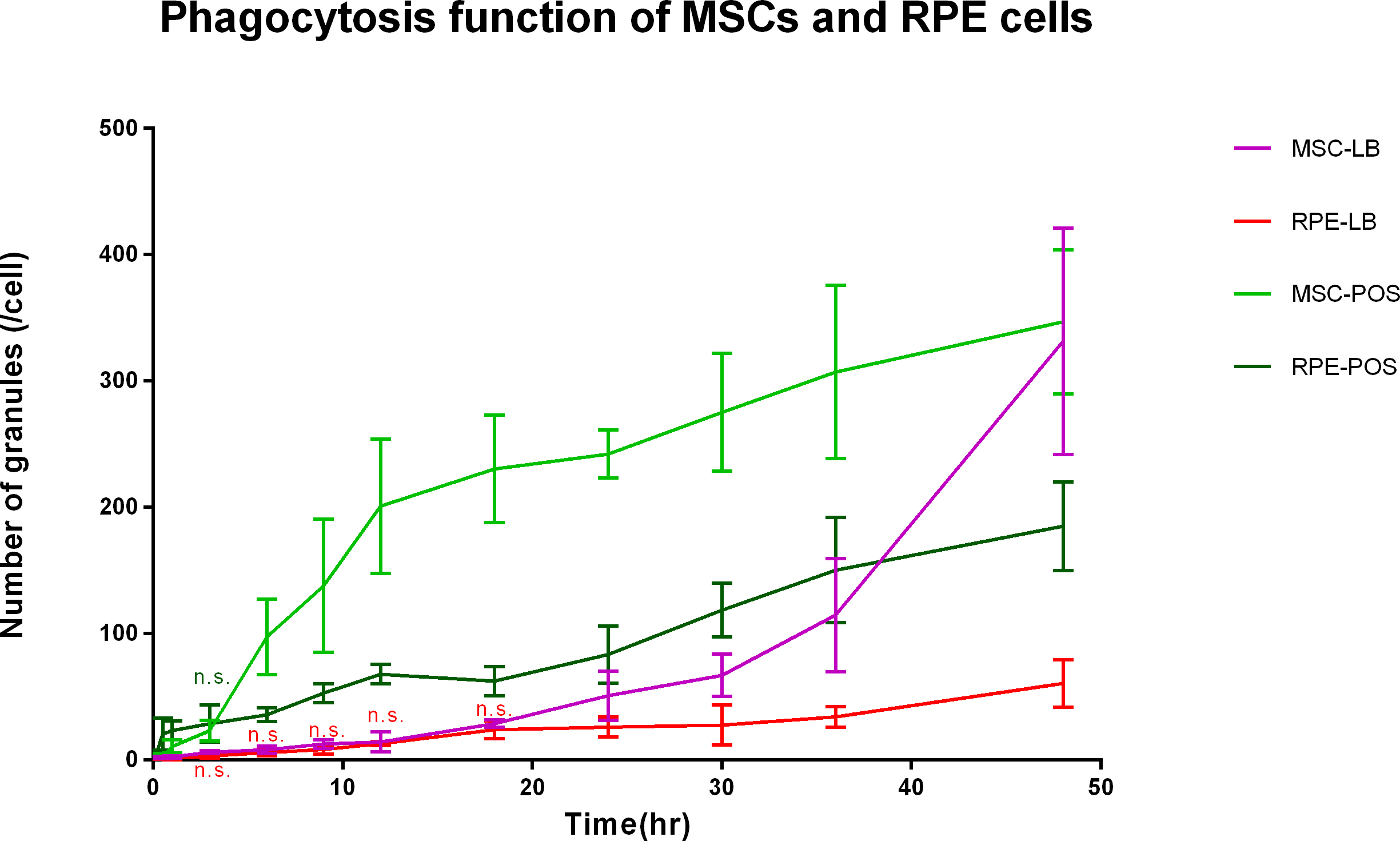
 Figure 5 of
Peng, Mol Vis 2017; 23:8-19.
Figure 5 of
Peng, Mol Vis 2017; 23:8-19.