Figure 4. POS phagocytosis by BM-MSCs and RPE cells. The first picture in rows 1 and 2 exhibits the photoreceptor outer segment (POS)
with fluorescent microscopy magnified 200X and 400X, respectively. Rows 1 and 3 show bone marrow mesenchymal stem cells (BM-MSCs)
incubated with the POS for different time periods. Rows 2 and 4 show RPE cells incubated with the POS for different time periods.
The POS phagocytized by the cells is yellow.
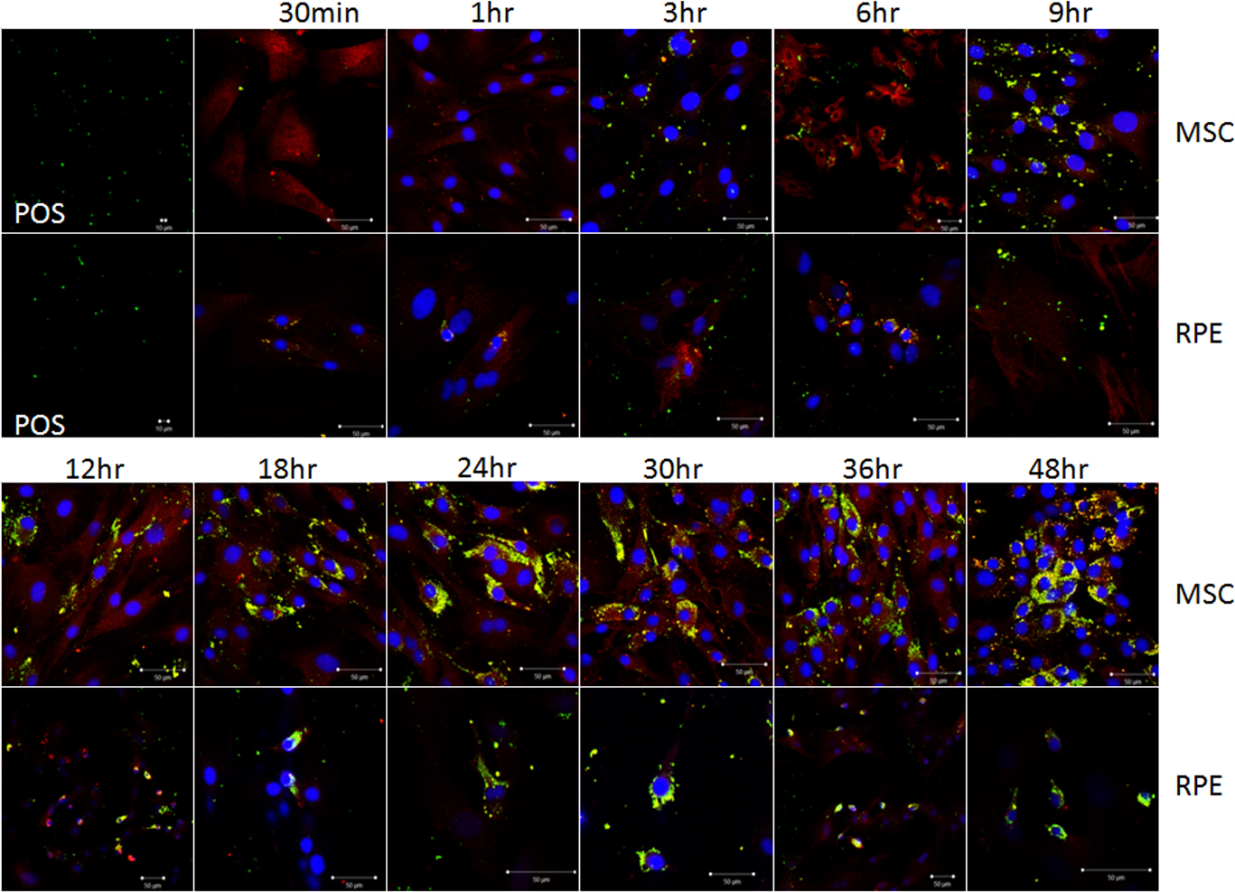
 Figure 4 of
Peng, Mol Vis 2017; 23:8-19.
Figure 4 of
Peng, Mol Vis 2017; 23:8-19.