Figure 1. Fundus and optical coherence tomography findings in a 17-year-old patient with heterozygous deletion in OTX2. Color fundus (A: Central fundus. B: Wide field imaging including the peripheral retina) images of the right eye show atypical maculopathy (A), and mid-peripheral pigmentation with mild arteriolar attenuation (B). C: Transfoveal optical coherence tomography scan shows intraretinal hyperreflective scar (blue arrow), intraretinal cyst-like
spaces (yellow arrow), RPE changes (red arrow), and foveal atrophy. The hyperreflective lesions may represent RPE hyperplasia
and migration; however, there is no sign of lipofuscin deposition such as that seen in macular pattern dystrophy.
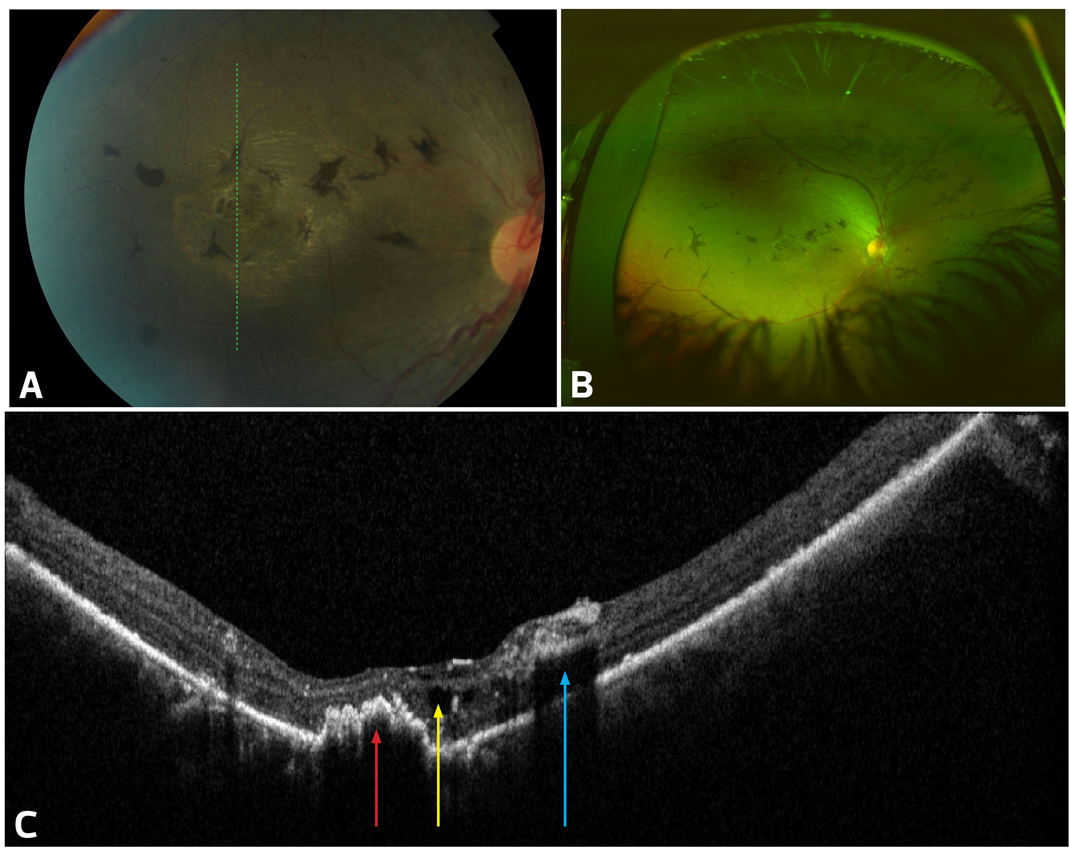
 Figure 1 of
Abdalla-Elsayed, Mol Vis 2017; 23:778-784.
Figure 1 of
Abdalla-Elsayed, Mol Vis 2017; 23:778-784.