Figure 5. Localization of CCL2 in the late stage of photic injury by double immunofluorescence staining. A, B, C: In the flat mount, CCL2 was colocalized in OX42-positive cells with amoeboid morphology at 3 days after light exposure.
D, E, F: At 7 days, no expression of CCL2 was observed in OX42-positive cells. G: However, expression of CCL2 was observed in cells with radially oriented processes throughout the inner nuclear layer. H: Strong immunoreactivity to glial fibrillary acidic protein (GFAP) appeared from the internal limiting membrane to the inner
plexiform layer at 7 days. I: The merged image shows that CCL2 was colocalized in GFAP-positive Müller glia. J, K, L: Amplified images of G, H, and I, respectively. M, N, O: Immunofluorescent staining in the retinal flat mount confirmed the observation in the retinal slice.
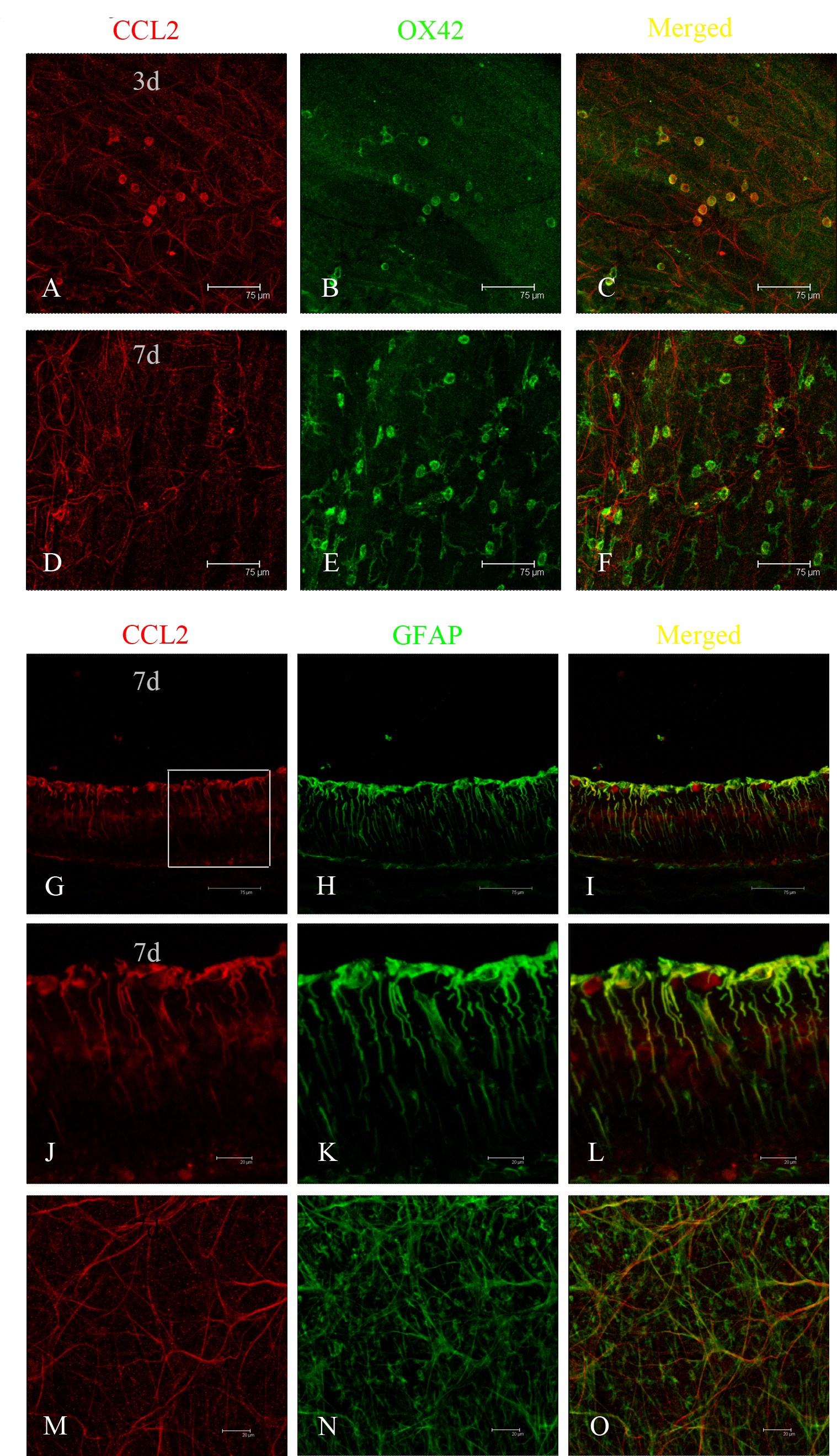
 Figure 5 of
Feng, Mol Vis 2017; 23:765-777.
Figure 5 of
Feng, Mol Vis 2017; 23:765-777.