Figure 4. Localization of CCL2 in activated microglia and monocytes by double immunofluorescence staining. A, B: At 3 days after light exposure, in the retinal slice, CCL2 expression was observed in the outer nuclear layer (ONL), and
many OX42-positive cells were activated and migrated into the ONL and subretinal space. C: The merged image shows that CCL2 was colocalized in those activated and migrated microglia and monocytes. D, E, F: Amplified images of A, B, and C, respectively. G, H: In the retinal flat mount, CCL2 was expressed richly in the outer retina, where OX42-positive cells were found. I: The merged image shows that CCL2 was colocalized in those microglia and monocytes. J, K, L: Amplified images of G, H, and I, respectively.
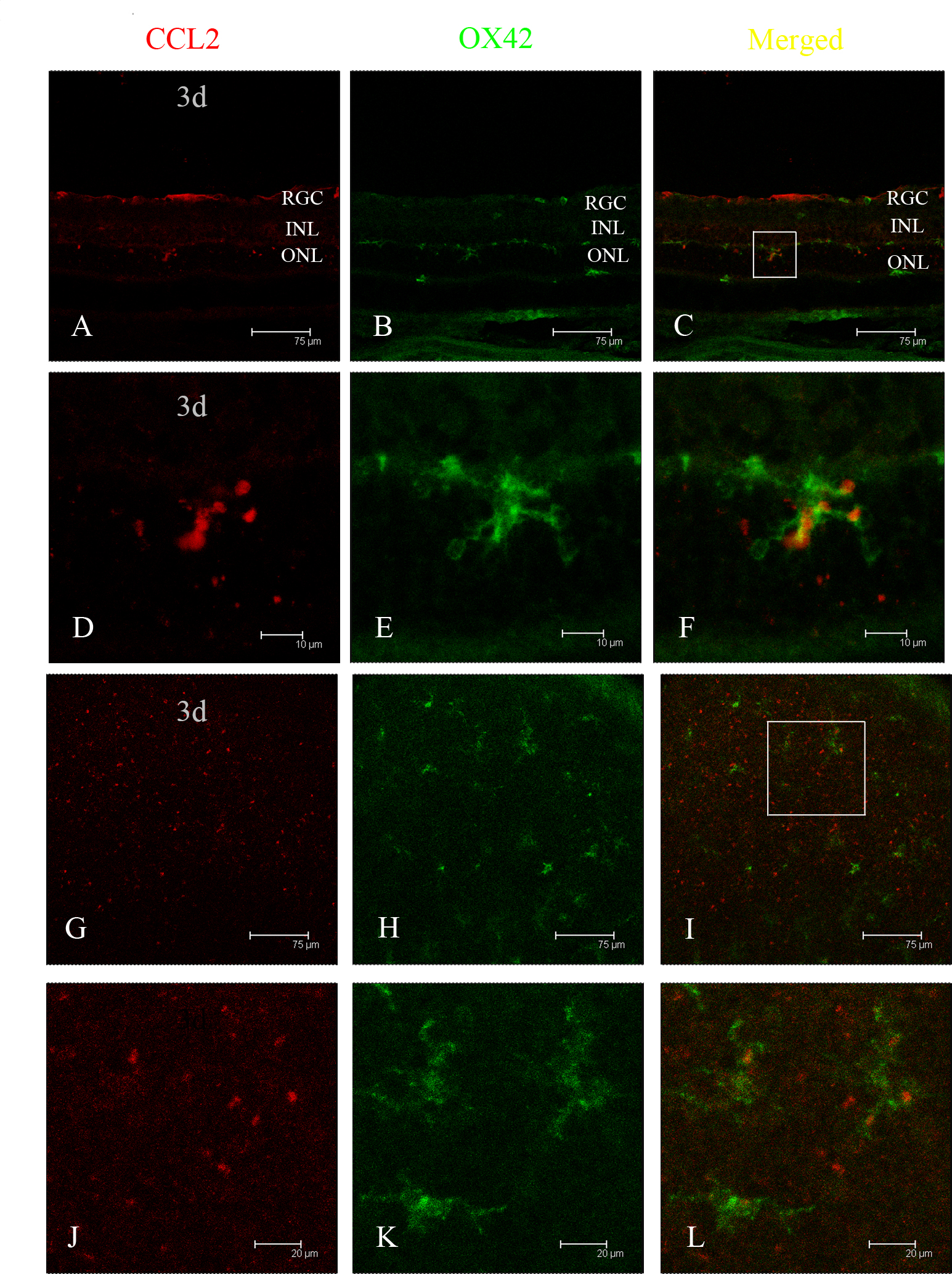
 Figure 4 of
Feng, Mol Vis 2017; 23:765-777.
Figure 4 of
Feng, Mol Vis 2017; 23:765-777.