Figure 3. Localization of CCL2 in a healthy retina and apoptotic photoreceptors by double immunofluorescence staining. A, D: In a healthy retinal slice and flat mount, CCL2 was mainly expressed in the retinal ganglion cell layer. B, E: Glial fibrillary acidic protein (GFAP) staining was localized in the astrocytes. C, F: The merged images revealed that CCL2 was expressed by astrocytes in a healthy retina. G: At 1 day after light exposure, CCL2 was expressed robustly in the outer nuclear layer (ONL). H: Large numbers of terminal deoxynucleotidyl transferase dUTP nick end labeling (TUNEL)–positive cells were observed in the
ONL. I: The merged image shows that some of the CCL2 expression was colocalized in TUNEL-positive cells. J, K, L: Amplified images of G, H, and I, respectively.
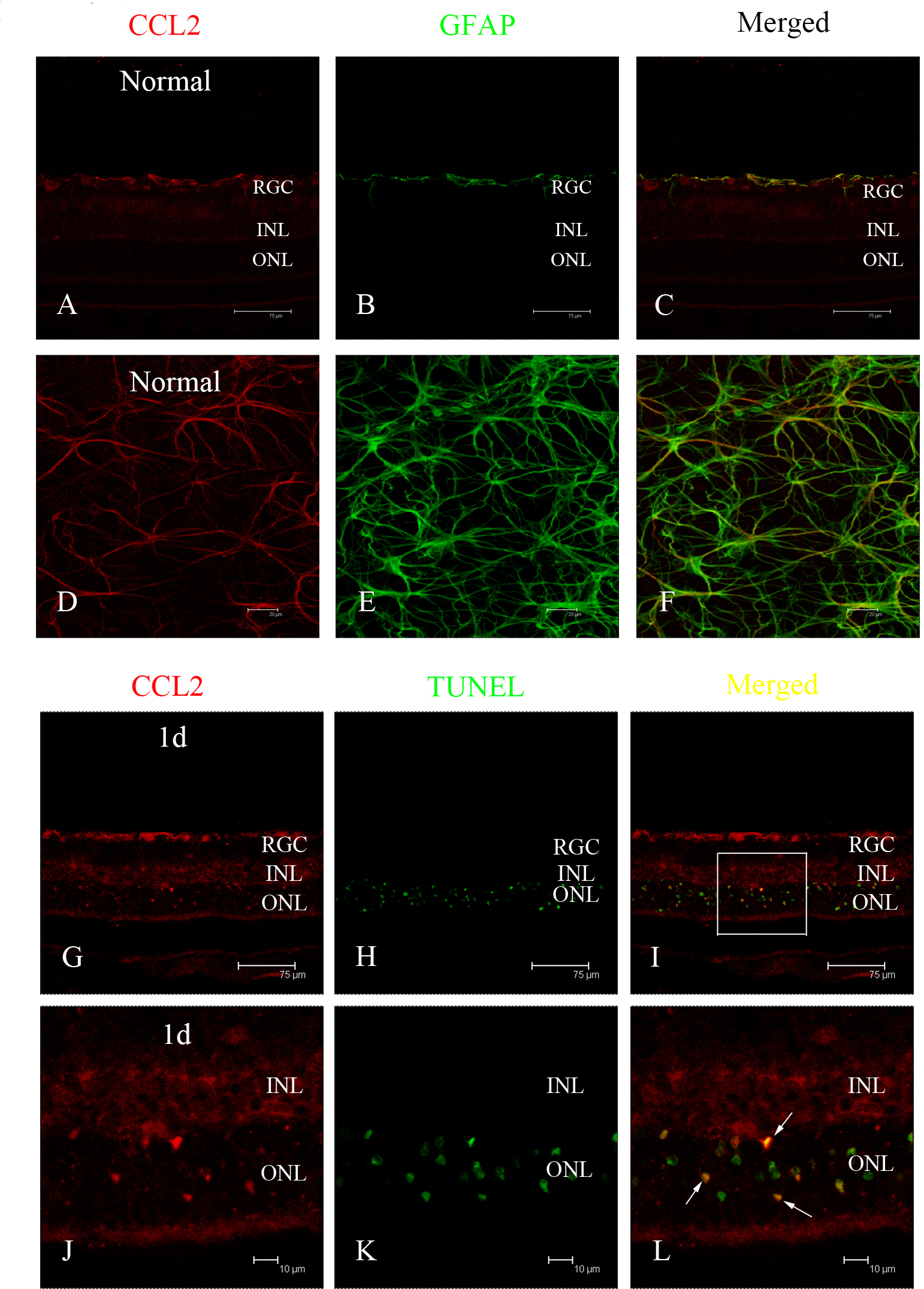
 Figure 3 of
Feng, Mol Vis 2017; 23:765-777.
Figure 3 of
Feng, Mol Vis 2017; 23:765-777.