Figure 1. Microglia/monocyte activation and infiltration in light-induced photoreceptor degeneration. A, E: In healthy retinas, OX42-positive microglia were seen only in the retinal ganglion cells and the inner plexiform layer with
long, slim processes, while no ED1-positive monocytes were seen in the retina. B, F: One day after exposure to light, a small number of monocytes/microglia began to infiltrate into the outer nuclear layer
(ONL) and subretinal space. C: The number of OX42-positive cells peaked at 3 days, with activated amoeboid morphology. D, H: At 7 days, many OX42-positive cells remained in the thinned ONL and subretinal space, while the number of ED1-positive cells
in the outer retina peaked at that time.
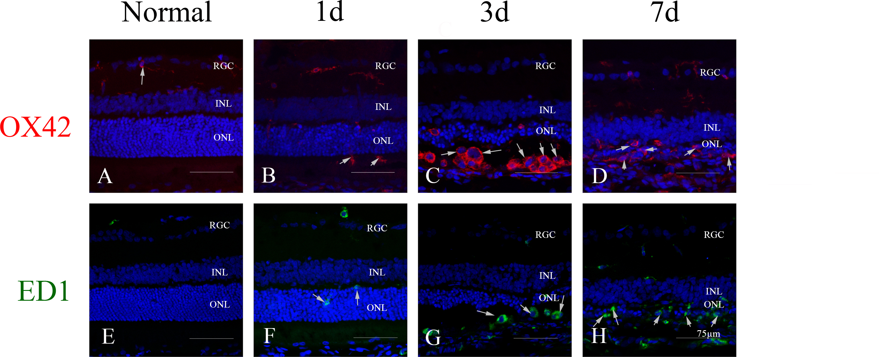
 Figure 1 of
Feng, Mol Vis 2017; 23:765-777.
Figure 1 of
Feng, Mol Vis 2017; 23:765-777.