Figure 5. Transmission electron micrographs of necrotic keratocytes in the experimental and control groups following 4 weeks of storage.
In the control lenticule, the chromatin in the cell nucleus was condensed, whereas in the lenticules in groups 1–8, there
was irregular clumping of chromatin, cytoplasmic vacuoles, and swelling nuclei. These necrotic keratocytes were less observed
in groups 7 and 8. Scale bar = 2 μm.
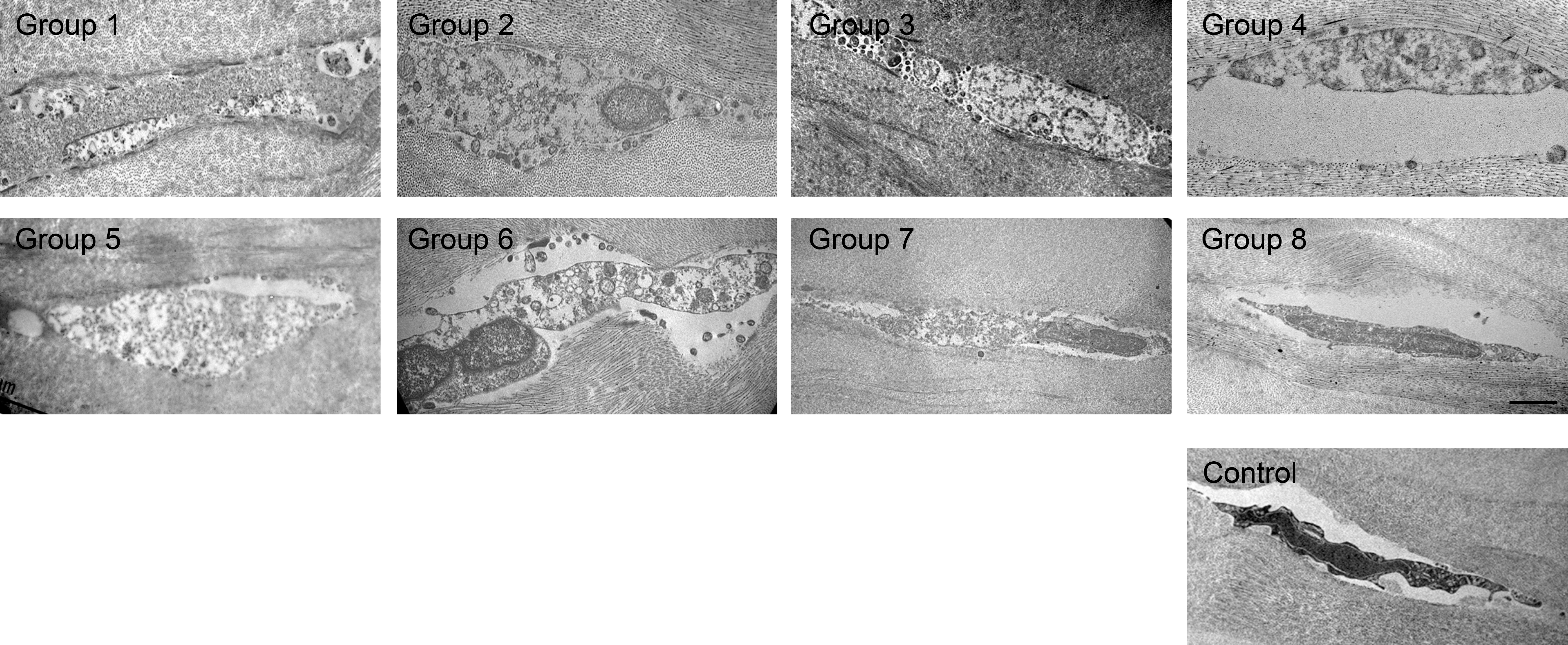
 Figure 5 of
Liu, Mol Vis 2017; 23:753-764.
Figure 5 of
Liu, Mol Vis 2017; 23:753-764.