Figure 3. Western blot analysis and retinal DNA levels in rats reared for 6 weeks in low- or high-cyclic light. Retinal proteins (20
μg/lane), pooled from four to five rats, were electrophoresed on 12.5% sodium dodecyl sulfate (SDS)-polyacrylamide gels, transferred
to polyvinylidene fluoride (PVDF) membranes, and then probed with antibodies as indicated in Appendix 1. Rats exposed to intense
light for 6 h and then kept in darkness for 2 weeks (B, E). The fellow eyes were used for DNA measurements (C), [n = 8 for No LD]. Density profiles determined by Image J analysis. A: Cone opsin levels were lower in all rats reared under high cyclic light than for those reared in low cyclic light, whereas
medium wavelength cone arrestin (mCAR) staining was lower in only the rats fed AREDS. There were no major differences in rhodopsin
and S-antigen levels for AREDS (A), zinc oxide (Z), or AREDS antioxidants minus zinc (AO). Multiple CEP protein adducts were
present, with greater staining for the 200 lux cyclic light-reared rats than for those reared in 20 lux light. B: Two weeks after exposure to acute intense light, higher levels of cone- and rod-opsins were present in rats from the high
light condition versus low cyclic light. S-antigen and mCAR levels were similar. C: Individual antioxidants had little effect on retinal DNA for rats reared in 20 lux cyclic light (55–79 μg/retina); DNA levels
for 200 lux cyclic light animals (107–128 μg) were significantly higher (p<0.05). D: Rats fed vehicle (V), rosemary (R), AREDS + rosemary (A+R), or zinc oxide + rosemary (Z+R). Cone opsin levels were higher
in rats reared in 20 lux cyclic light than for those reared in 200 lux light. Rhodopsin (monomer) and S-antigen levels were
unchanged by higher light rearing conditions, or by antioxidant feeding E: Two weeks after photooxidative damage considerable loss of cone opsin and rhodopsin (monomer) occurred in rats originally
from the 20 lux light environment, and rhodopsin was still undergoing degradation. mCAR levels were preserved in A+R and Z+R
rats, whereas S-antigen levels were low in all retinas. Glyceraldehyde phosphate dehydrogenase (GAPDH) was the control for
protein loading.
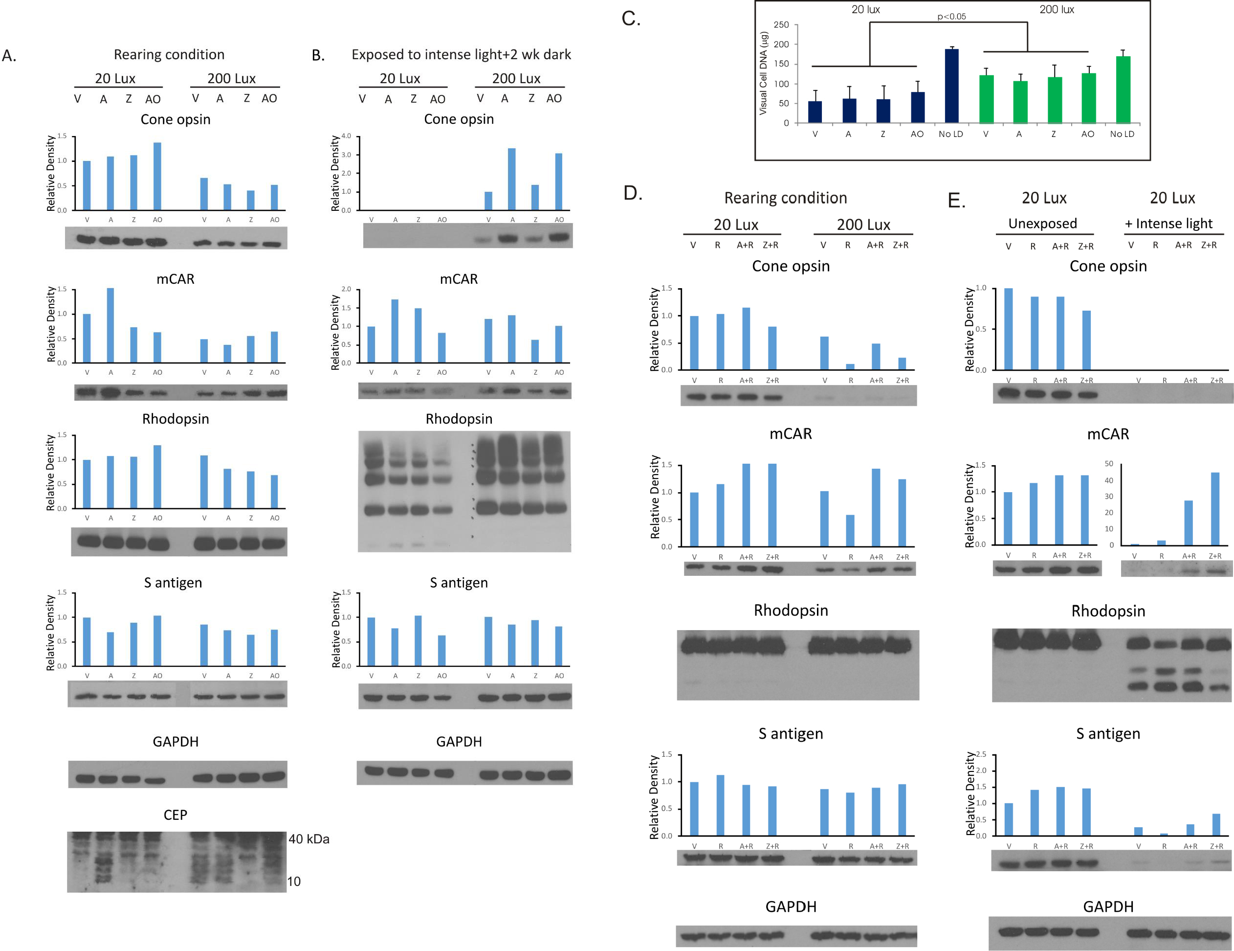
 Figure 3 of
Wong, Mol Vis 2017; 23:718-739.
Figure 3 of
Wong, Mol Vis 2017; 23:718-739.