Figure 2. Confirmation of differential sex- and age-related retina gene expression with qPCR. Quantitative PCR (qPCR) analysis of selected
targets identified in the microarray study confirmed differentially expressed retina genes that are associated with aging
in female and male animals (A), sexually dimorphic aging changes (B), and changes related to sex differences (C). ND = not detectable. Data are shown as fold changes relative to young male animals for the microarray and qPCR data. Box
plots denote median values with 25–75th percentiles quartiles, and whiskers define the 10–90th percentiles; n = 7–8 samples per group, for a total number of 47 samples analyzed. P values were determined with two-way
ANOVA (age × sex), followed by the Student–Newman–Keuls post-hoc test. *p<0.05, **p<0.01, ***p<0.001. Solid lines denote comparisons
of age-related changes within a sex group, and dashed lines are comparisons of sex-related differences within an age group.
Cx3cr1 = chemokine (C-X3-C motif) receptor 1; Rdh9 = retinol dehydrogenase 9; Edn2 = endothelin 2; Ccl21b = chemokine (C-C
motif) ligand 21B; Lcn2 = lipocalin 2; Gbp4 = guanylate binding protein 4; Rgr = retinal G protein coupled receptor; Ddx3y
= DEAD (Asp-Glu-Ala-Asp) box polypeptide 3, Y-linked; Rtn4 = reticulon 4; Hes5 = hairy and enhancer of split 5.
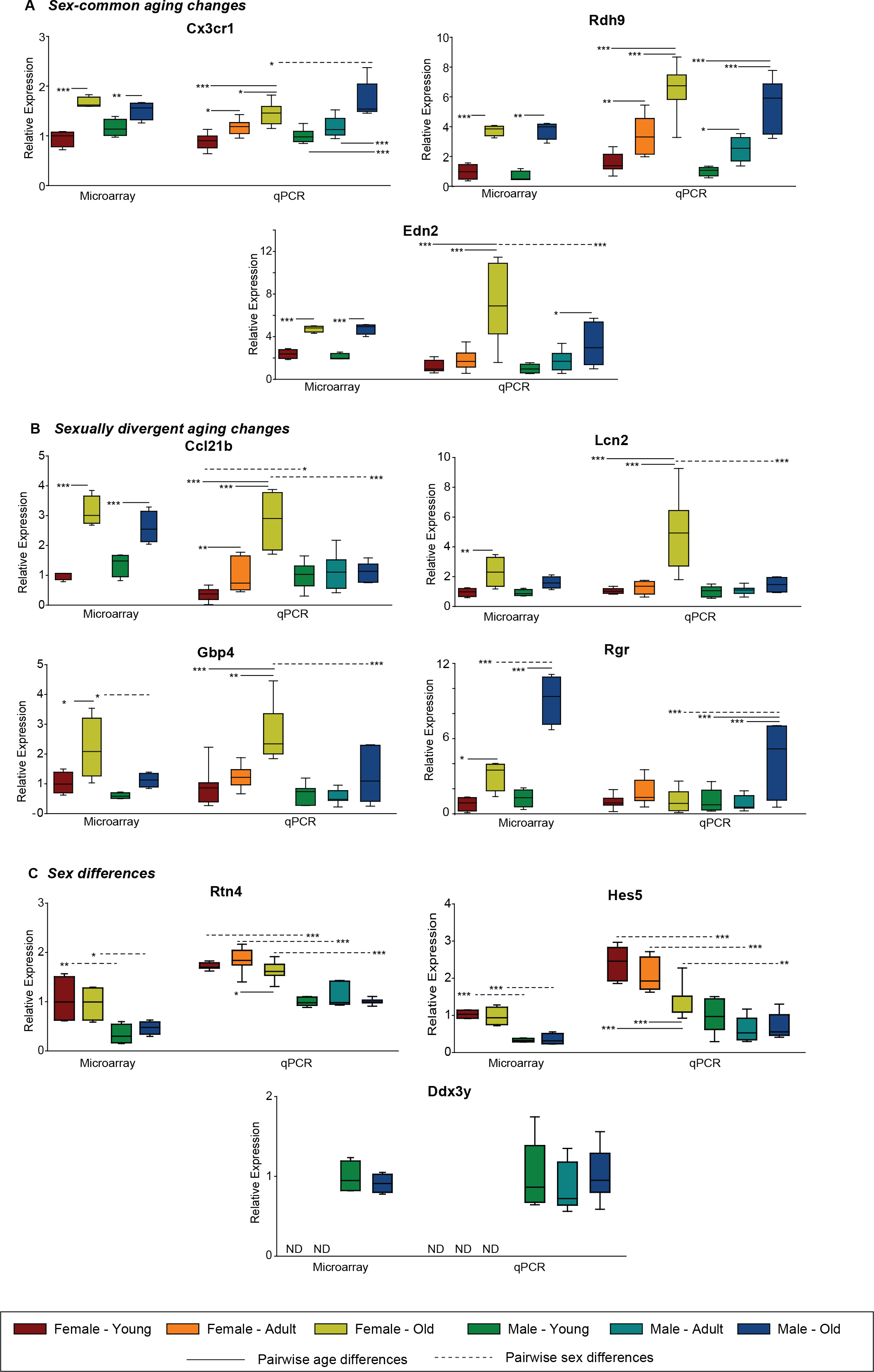
 Figure 2 of
Du, Mol Vis 2017; 23:707-717.
Figure 2 of
Du, Mol Vis 2017; 23:707-717.