Figure 1. Exam findings for Patient 1. A: Full-field flash electroretinography according to International Society for Clinical Electrophysiology of Vision (ISCEV)
standards is shown at ages 3.5 months (black traces) and 7.5 months (gray traces). At 3.5 months, the electroretinogram (ERG)
traces showed notably decreased amplitudes in the rod and cone responses, with the amplitudes in the rod-specific, maximal
combined rod-cone, and cone-specific responses at 10% of normal. No obvious electronegative aspect was evident at that time.
At 7.5 months, the ERG traces showed significantly reduced amplitudes in the rod and cone responses and an electronegative
waveform on maximal responses. Control ERGs performed at the same institution for a healthy 8-month-old are included for comparison
(red traces). DA = dark adapted; LA = light adapted. B: Right and left fundus photographs showing essentially normal fundi at age 4 years. C: Family pedigree of Patient 1.
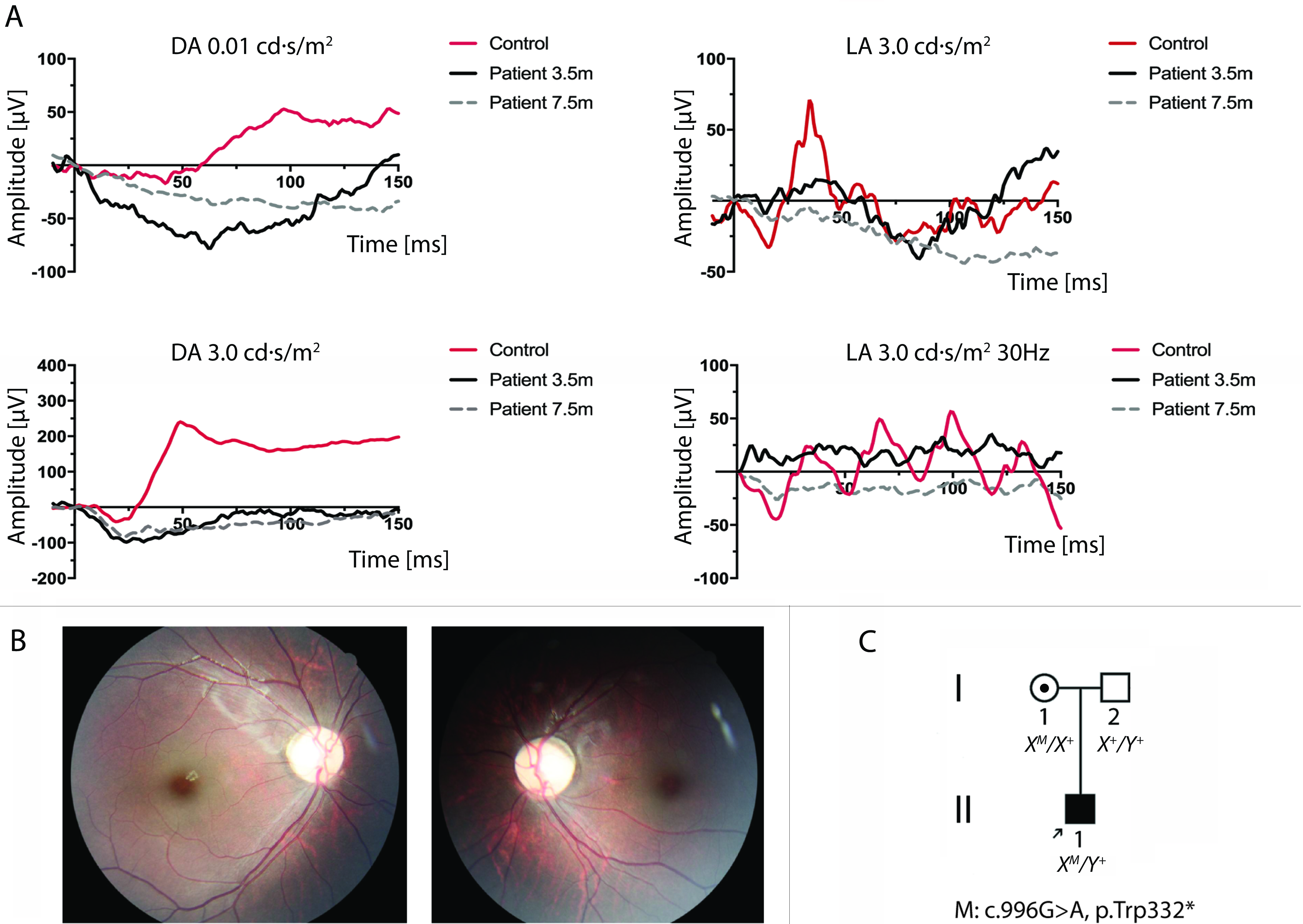
 Figure 1 of
Men, Mol Vis 2017; 23:695-706.
Figure 1 of
Men, Mol Vis 2017; 23:695-706.