Figure 6. Immunohistochemistry analysis of uveal melanoma multicellular tumor spheroids shows positive staining for SSX4 (green), Hoechst
staining of nucleus (blue; A) with corresponding light-microscopic image (B). The presence of synovial sarcoma X breakpoint protein 4 (SSX4) was verified with RNAscope staining (red), Hoechst staining
of nucleus (blue; C) where SSX4 RNA transcripts are shown as red chromogenic dots, and with the corresponding light-microscopic image (D). E: Percentage of SSX4-positive cells in multicellular tumor spheroids (MCTS) (D1, D2, and D3) versus uncultured primary tumors
(D1, D2, and D3) analyzed with immunohistochemistry (IHC).
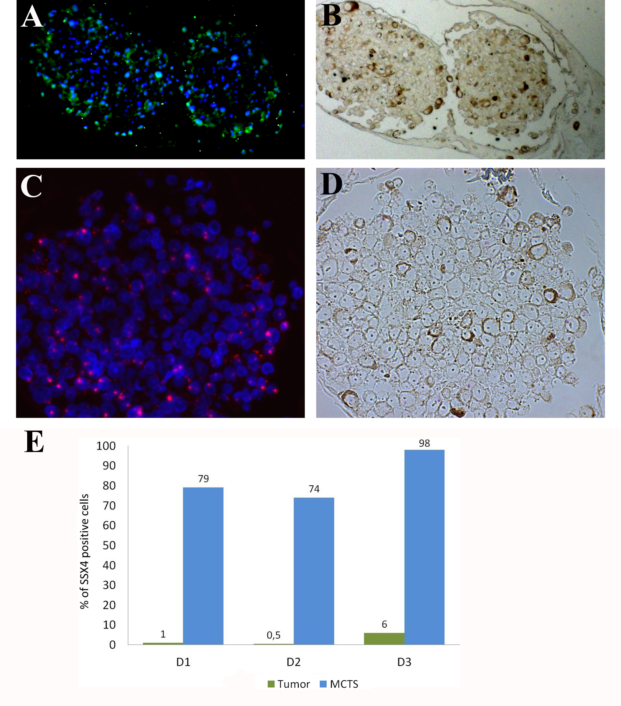
 Figure 6 of
Ness, Mol Vis 2017; 23:680-694.
Figure 6 of
Ness, Mol Vis 2017; 23:680-694.