Figure 1. Multicellular tumor spheroid culture of primary uveal melanoma cells. A: Single cells (upper inset) after primary tumor isolation, during cultivation small pigmented tumor spheres formed (lower
inset), and further developing resulting into large spheroid structures if not passaged. B: Adherent cell culture of primary uveal melanoma (UM) cells. C: Ki67 staining (*) of UM multicellular tumor spheroid (MCTS). D: Immunohistochemical staining of antimelanoma (green) and Hoechst staining of the nucleus (blue; right panel) with the corresponding
light-microscopic image (left panel) of UM MCTS.
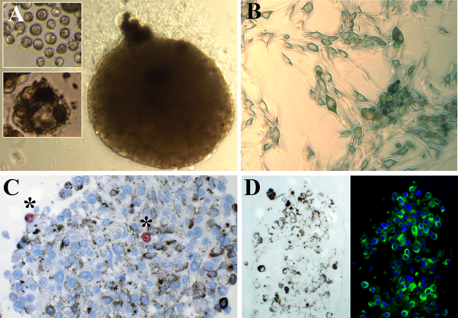
 Figure 1 of
Ness, Mol Vis 2017; 23:680-694.
Figure 1 of
Ness, Mol Vis 2017; 23:680-694.