Figure 4. Thermal denaturation of WT and MU βB1 monomers and heteromers with αA-crystallin. Thermal denaturation curves were obtained
by heating 0.2 mg/ml of the wild-type (WT) and mutant (MU) βB1 proteins at 55 °C in a 50 mM phosphate buffer, pH 7.4, and
then measuring light scattering at 405 nm. Meanwhile, the WT βB1 protein was heated at 55 °C with an equal molar amount of
αA-crystallin, and the results were compared with those for heating the MU βB1 protein with an equal molar amount of αA-crystallin.
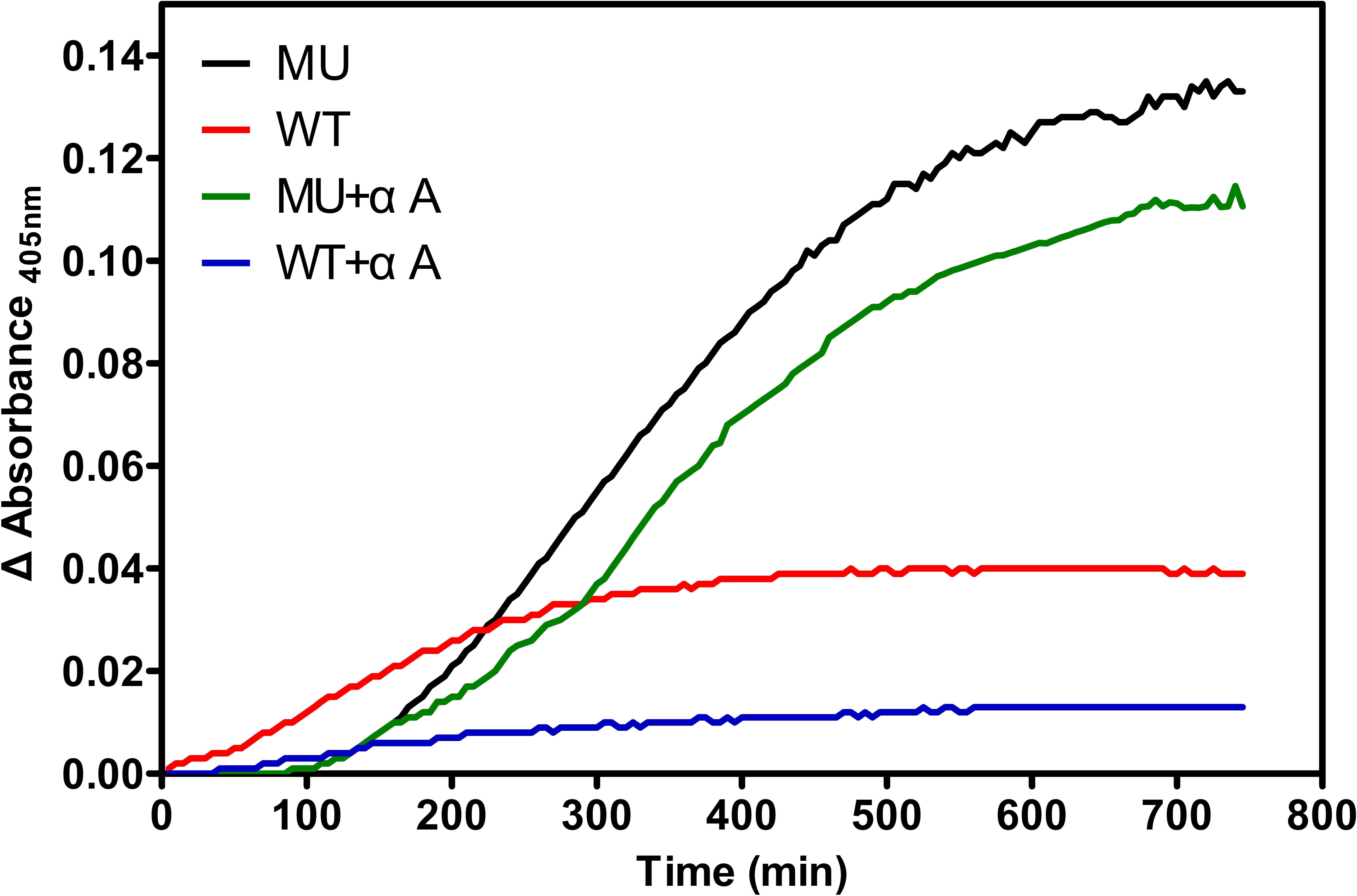
 Figure 4 of
Rao, Mol Vis 2017; 23:624-637.
Figure 4 of
Rao, Mol Vis 2017; 23:624-637.