Figure 3. Clinical examinations of the patients with mild FEVR. A: Fundus image of the proband with mild familial exudative vitreoretinopathy (FEVR). B: Fundus fluorescein angiography (FFA) shows the vessels in the peripheral retina walking as broom-like, and the temporal
peripheral retinal shows capillary nonperfusion zones. C: Spectral domain optical coherence tomography (SD-OCT) detected retinal dystrophy and a hyperreflective epiretinal membrane-like
appearance in the inner surface of the retina.
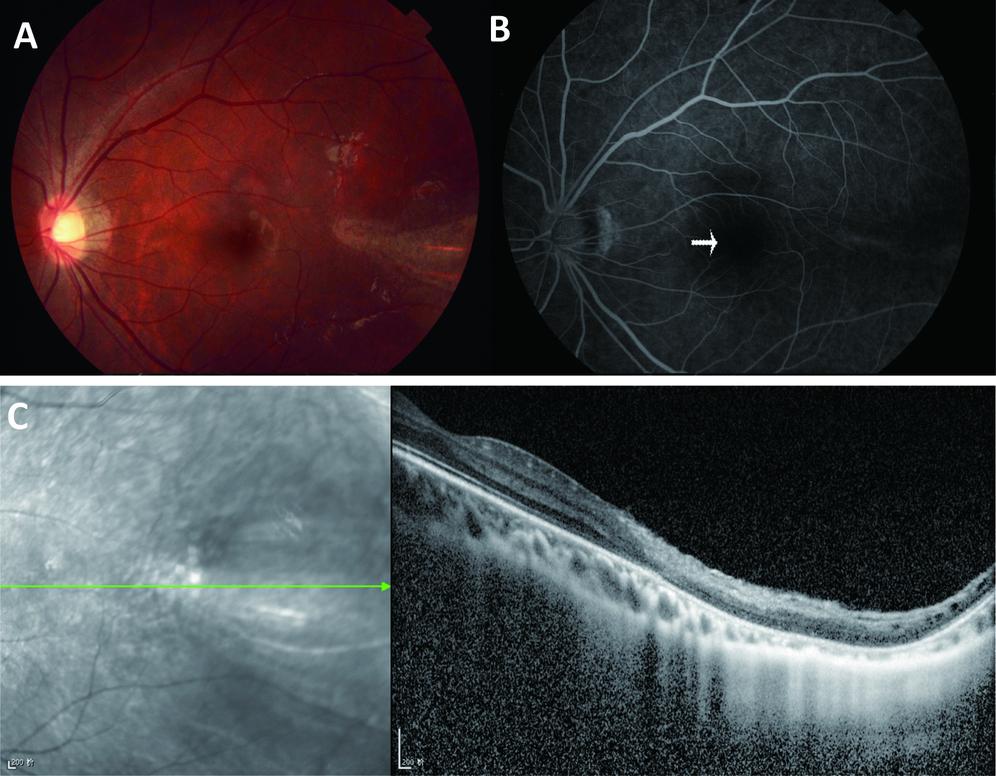
 Figure 3 of
Huang, Mol Vis 2017; 23:605-613.
Figure 3 of
Huang, Mol Vis 2017; 23:605-613.