Figure 2. Clinical examinations of patients with severe FEVR. A: B-scan ultrasonographic of the proband in family A reveals an irregular peripheral wall of the eyeball and persistent fetal
vasculature with the hyaloid artery. B: Fundus image of the proband in family A. C: The same for family B. D: Spectral domain optical coherence tomography (SD-OCT) detected retinal detachment and hyperreflective epiretinal membrane-like
appearance in the inner surface of the retina. E: Fundus fluorescein angiography (FFA) shows the various vascular branches of the posterior pole retina. The temporal peripheral
retinal shows nonperfusion zones along with abnormal new blood vessels and fluorescence leakage.
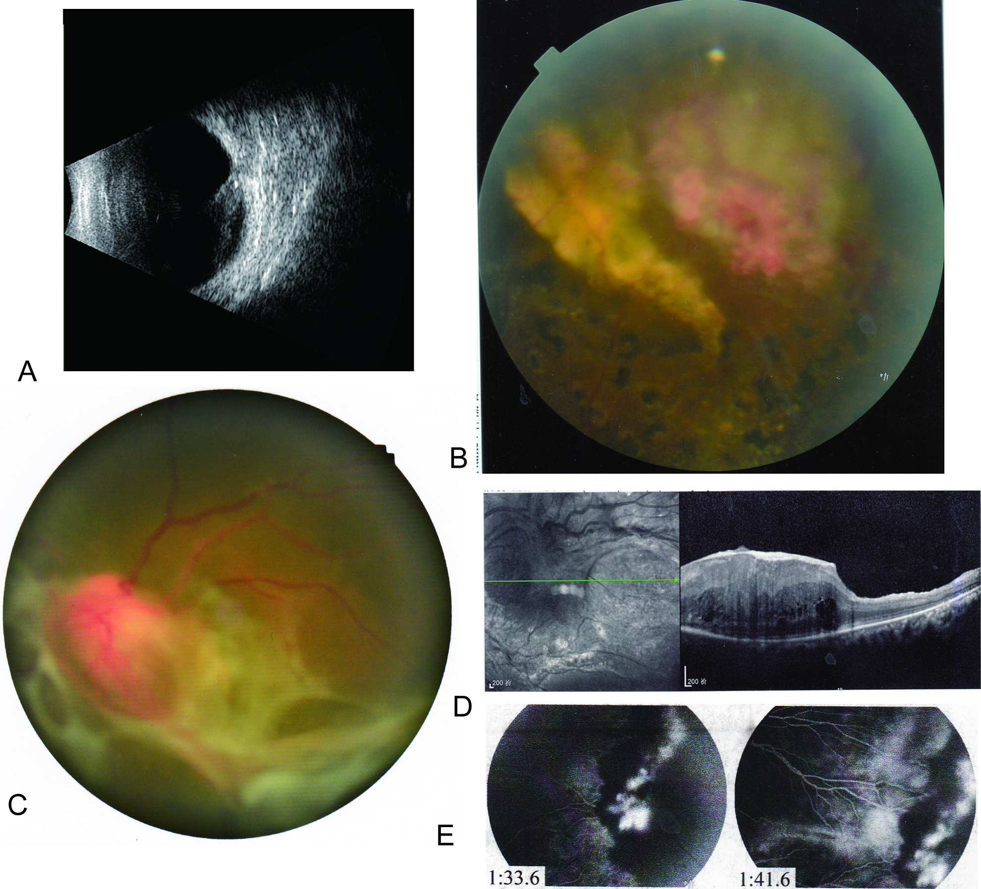
 Figure 2 of
Huang, Mol Vis 2017; 23:605-613.
Figure 2 of
Huang, Mol Vis 2017; 23:605-613.