Figure 6. Differentiation of RPE cells increases non-visual cycle gene expression. ARPE-19 cells grown for either 4 days or 4 months
were used for total RNA and protein extractions and were used for real-time quantitative PCR and western blotting, respectively,
as described in the Methods section. A: Real-time PCR analysis of RPE-specific non-visual cycle mRNA expression. The values are mean ± standard deviation (SD),
n = 4. *p<0.001 compared with control. B: Western blot analysis of the expression of the RPE-specific non-visual cycle proteins. α-Tubulin expression shows that the
amount of protein used in different samples is similar.
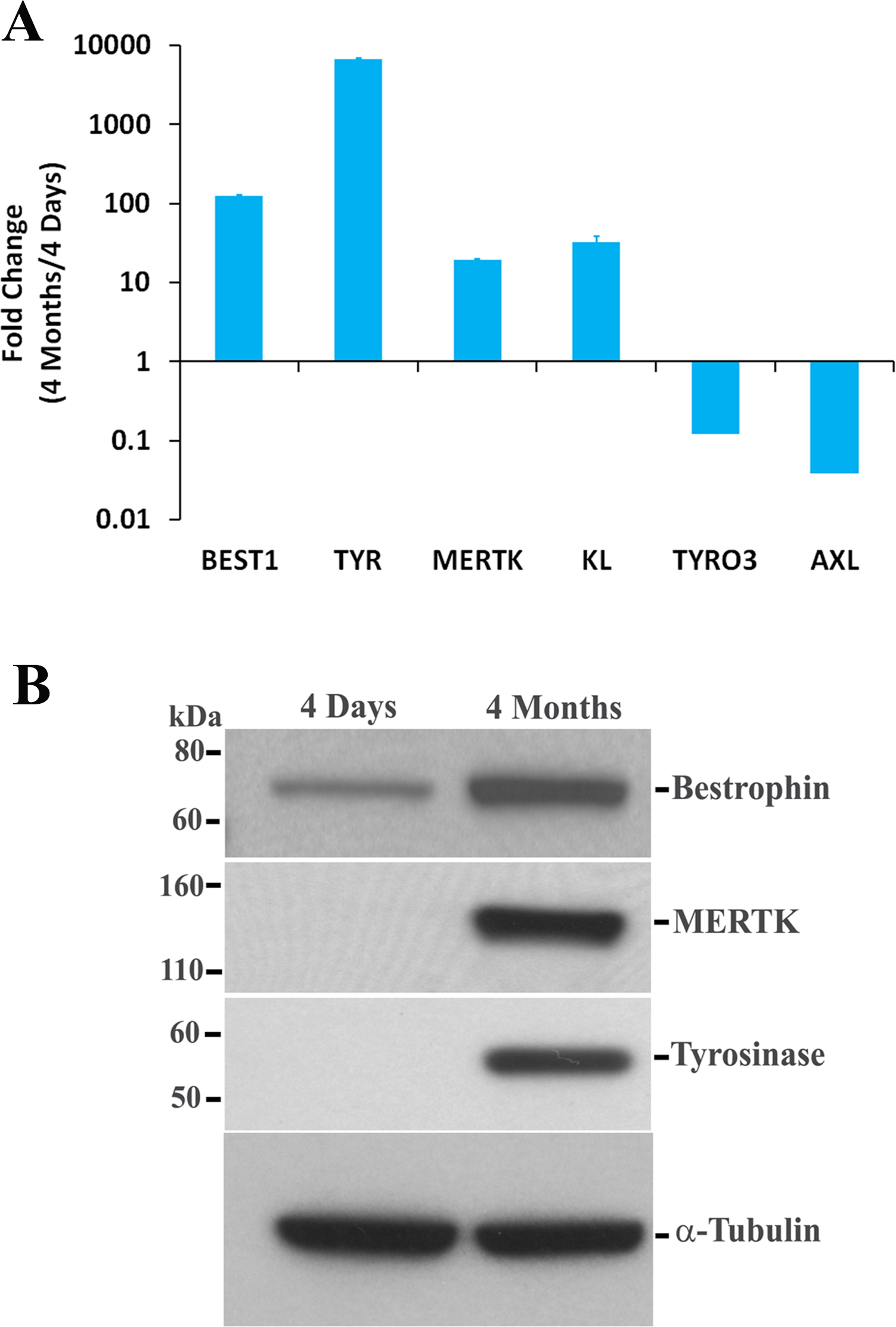
 Figure 6 of
Samuel, Mol Vis 2017; 23:60-89.
Figure 6 of
Samuel, Mol Vis 2017; 23:60-89.