Figure 2. Confocal micrographs of ARPE-19 cells cultured for 4 months. The ARPE-19 cells were plated on laminin-coated Transwell® filter membrane at a density of 3 × 10⁵ cells/cm2 and grown for 4 months as outlined in the Methods section. Rabbit anti- zonula occludens-1 (ZO-1), mouse anti-premelanosome
protein (PMEL), mouse anti-claudin-2 primary antibodies, and Alexa Fluor secondary antibodies were used to immunostain the
ARPE-19 cells at 4 months post-confluency; rhodamine-phalloidin was used to stain actin. A: PMEL immunostaining correlates with the presence of melanosome pigmentation. B: ZO-1 immunostaining shows good junctional development and localization at the cell borders containing a mixture of elongated
and polygonal cells. C: Actin immunostaining reveals circumferential actin distribution throughout the height of the cells. D: Claudin-2 immunostaining shows tight junction localization uniformly across the monolayer. E: ZO-1 immunostaining shows good junctional development and localization at the cell borders regardless of the secondary antibody
used. F: Claudin-2 immunofluorescent labeling shows colocalization with ZO-1 in the tight junction complexes (merged image of D and E). Images were taken using an Andor Revolution XD spinning disk confocal microscope using the 40X Plan Fluor oil immersion
objective. Scale bar = 15 μm.
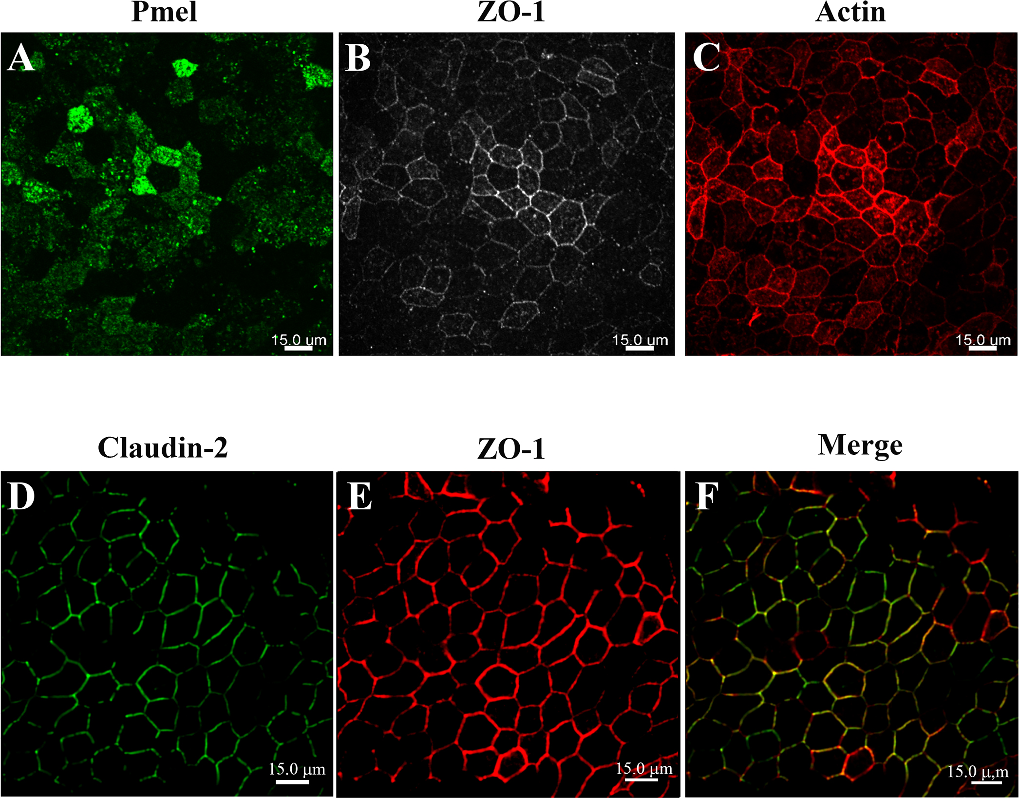
 Figure 2 of
Samuel, Mol Vis 2017; 23:60-89.
Figure 2 of
Samuel, Mol Vis 2017; 23:60-89.