Figure 2. Comparisons of extraocular muscles among KO and HT mice. Selected coronal image slices for four mice: two KO (knockout) and
two HT (wild-type) mice. The image slice is arranged from posterior to anterior when the slice number increases from small
to large. The red lines are the automatic segmentation results using in-house software. The in-pane resolution is 1.0 by 1.0
mm2, and the slice thickness is 1.0 mm.
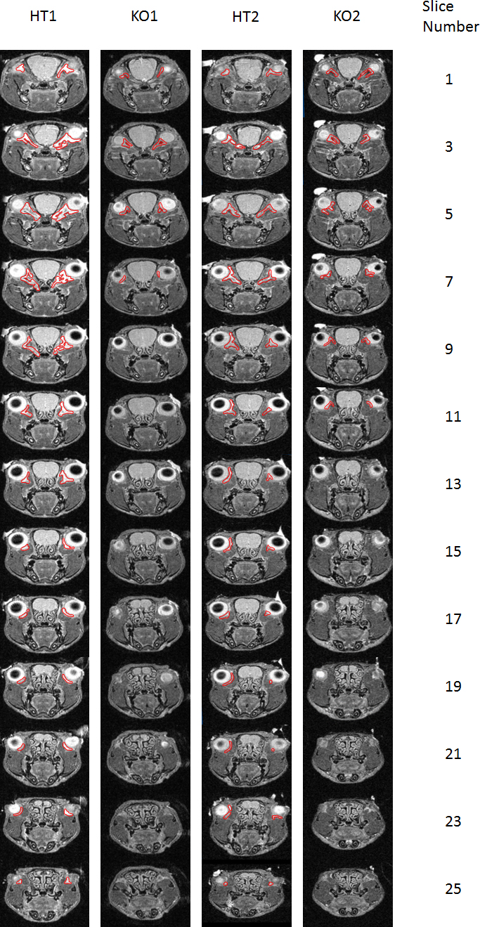
 Figure 2 of
Lee, Mol Vis 2017; 23:572-578.
Figure 2 of
Lee, Mol Vis 2017; 23:572-578.