Figure 2. Optic disc and Humphrey visual field deviations in patients with and without mutations in OPA1. A, B: Selected examples of fundus photographs of optic discs demonstrating temporal pallor (top) and corresponding Humphrey visual
field (HVF) pattern standard plots (bottom) in two patients with optic atrophy: one without an OPA1 variant (or other known gene; A), and one with a deletion extending from exon 1 to 28 (B). The HVF pattern standard plots show bilateral central/paracentral, temporal, and inferior Bjerrum defects for the mutation
negative patient (A) and bilateral centrocecal defects for the mutation positive patient (B).
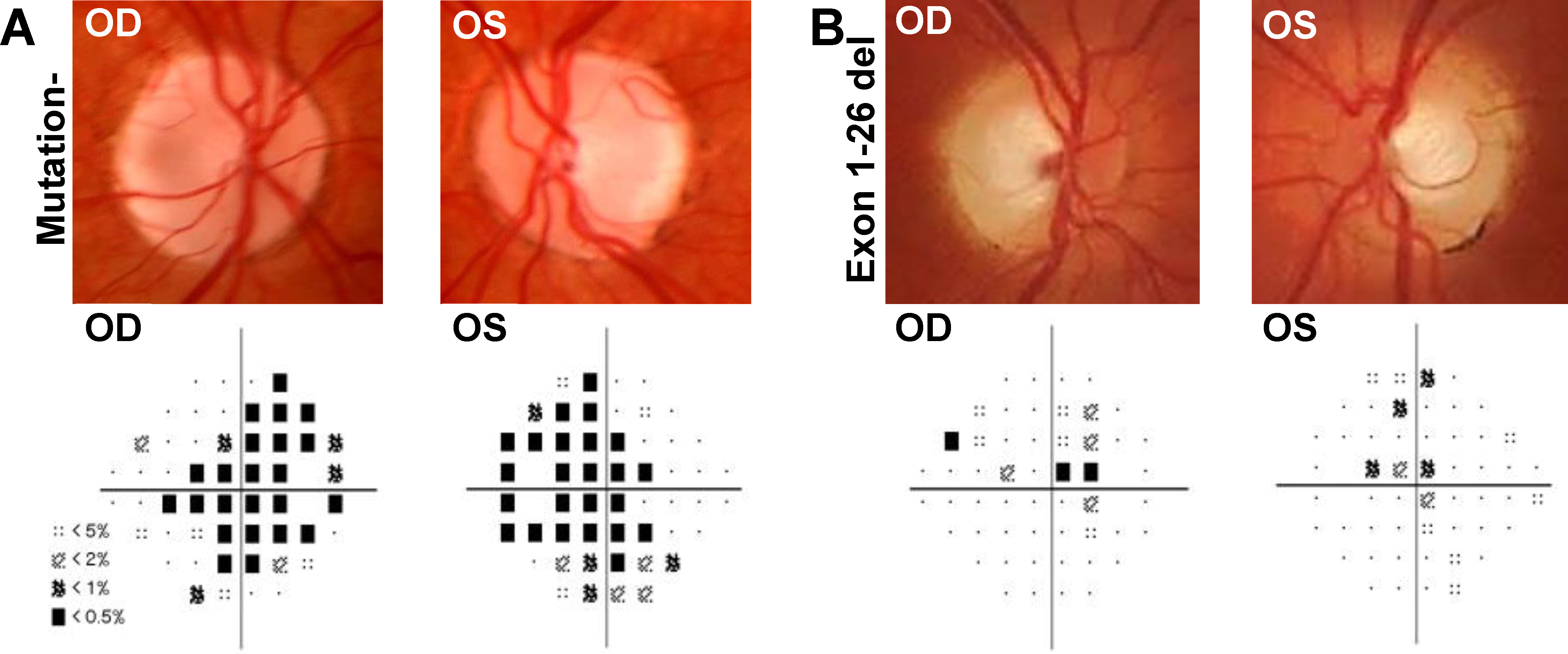
 Figure 2 of
Gaier, Mol Vis 2017; 23:548-560.
Figure 2 of
Gaier, Mol Vis 2017; 23:548-560.