Figure 1. Taxifolin prevented the decrease in retinal pigment epithelial (RPE) cell viability induced by H2O2. A: ARPE-19 cells were incubated with different concentrations of taxifolin (0, 10, 20, 50, and 100 µg/ml) for 24 h. B: ARPE-19 cells were treated with different concentrations of H2O2 (0, 50, 100, 200, and 400 µM) for 24 h. C: ARPE-19 cells were treated with 0.4 mM H2O2 for 24 h in the presence of different concentrations of taxifolin. Data are shown as the mean ± SD (n = 3); *p<0.05, **p<0.01,
and *** p<0.001.
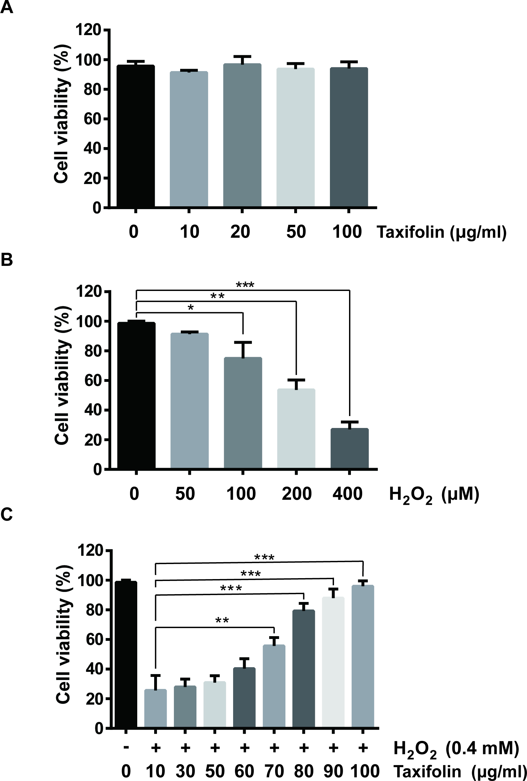
 Figure 1 of
Xie, Mol Vis 2017; 23:520-528.
Figure 1 of
Xie, Mol Vis 2017; 23:520-528.