Figure 7 of
Song, Mol Vis 2017; 23:504-513.
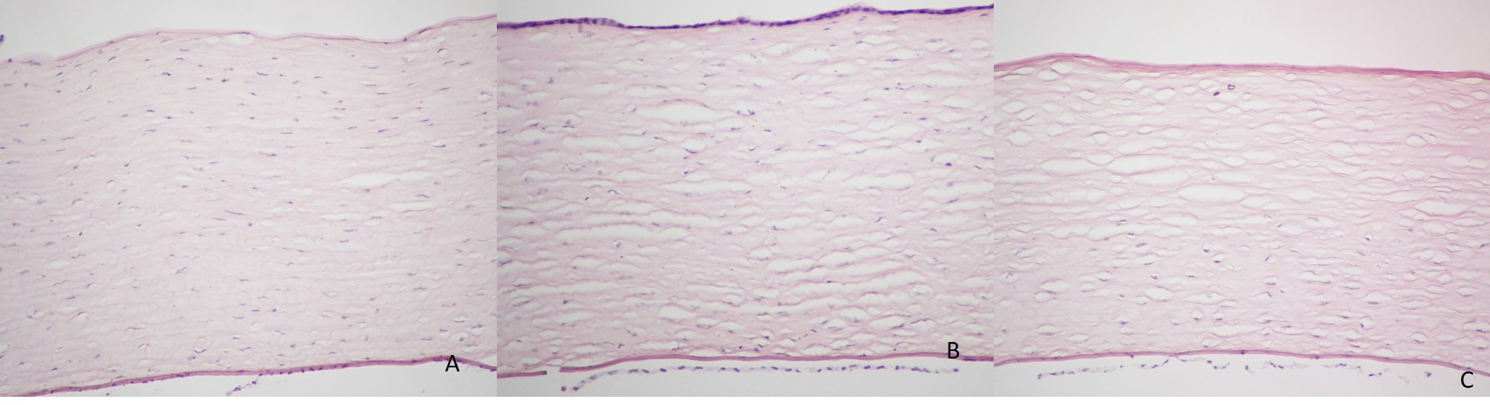
Figure 7.
Representative images of the HE stain. In the PBS group (
A
) and the GP-CXL group (
B
), the corneal structures are normal (200X).
C
: In the UVA-CXL group, keratocytes are absent in the anterior and middle stroma.
 Figure 7 of
Song, Mol Vis 2017; 23:504-513.
Figure 7 of
Song, Mol Vis 2017; 23:504-513.