Figure 6. Representative images from TEM of keratocytes after surgery (10,000X). A: In the PBS group, the keratocyte structures are normal. B: In the 0.2% GP-CXL group, the keratocyte structures are normal. There is no difference between the GP-CXL and PBS groups.
In the UVA-CXL group, the keratocytes are absent in the anterior and middle stroma. C: Only residual traces and apoptotic bodies (black triangle) are seen. D: In the posterior stroma, the apoptotic keratocytes
show typical features of apoptotic chromatin condensation (black star).
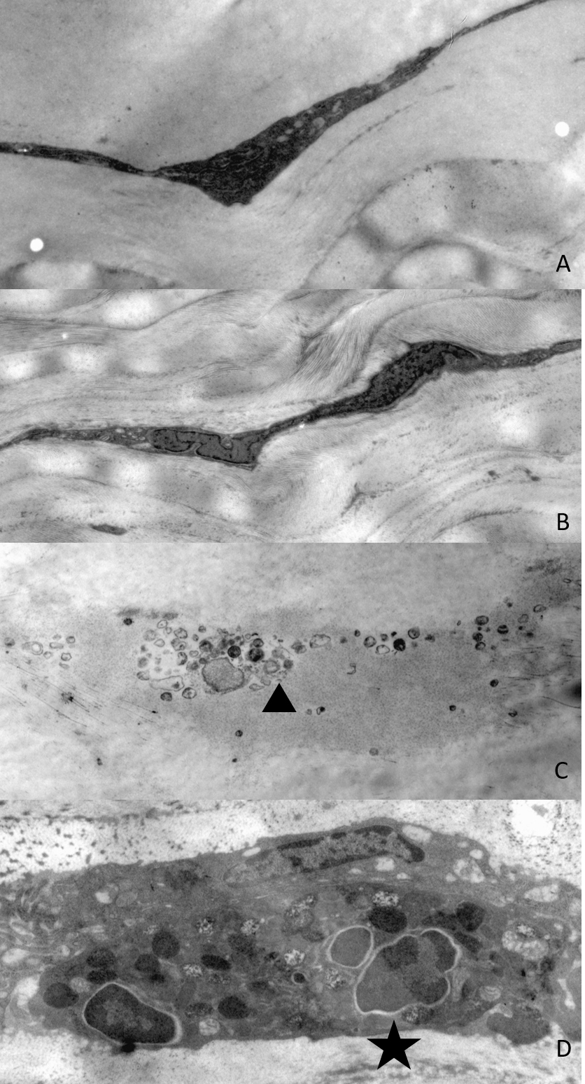
 Figure 6 of
Song, Mol Vis 2017; 23:504-513.
Figure 6 of
Song, Mol Vis 2017; 23:504-513.