Figure 4. Representative SEM images of endothelial cells after surgery (1,000X). A: Endothelial cells in the PBS group appear the same as endothelial cells in the 0.2% GP-CXL group. B: In the GP-CXL group, solitary cell damage can be seen, with the remaining endothelial cells maintaining a hexagonal structure.
C: In the UVA-CXL group, substantial areas of endothelial damage are seen. The damaged endothelium contains many small cavities
(black triangle) resembling holes in a sponge, and the normal hexagonal structure is absent.
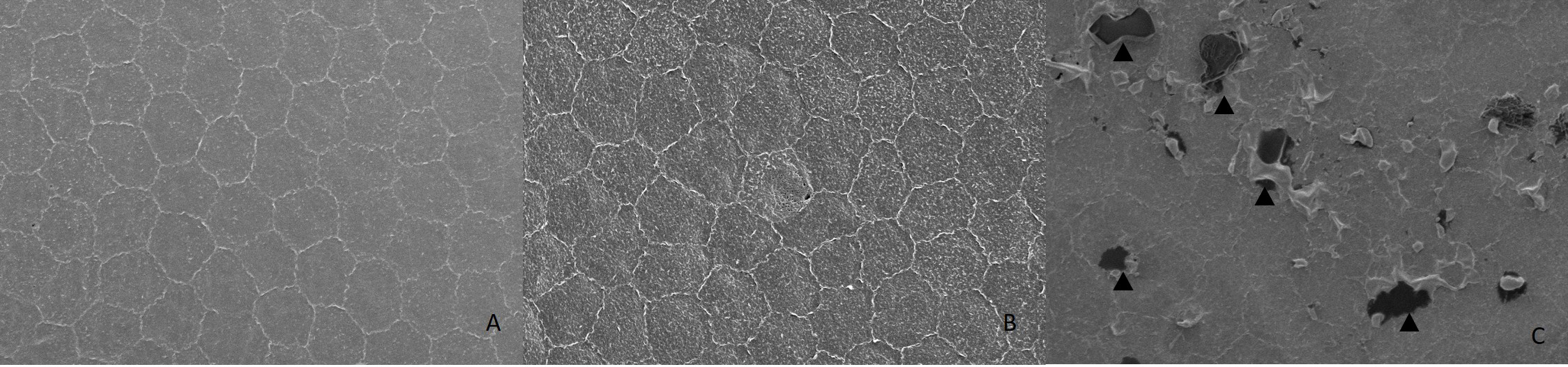
 Figure 4 of
Song, Mol Vis 2017; 23:504-513.
Figure 4 of
Song, Mol Vis 2017; 23:504-513.