Figure 3. Alizarin red and trypan blue staining of the endothelium after surgery. A: The cells are seen as complete endothelial cells in the PBS group. B: The endothelial cell borders in the 0.2% GP-CXL group are clear. C: In the UVA-CXL group, many apoptotic cells are present.
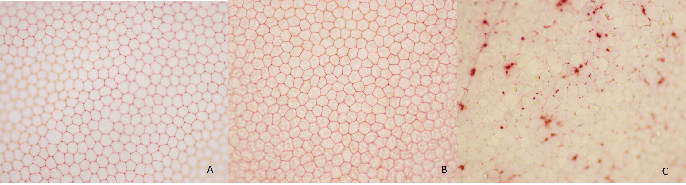
 Figure 3 of
Song, Mol Vis 2017; 23:504-513.
Figure 3 of
Song, Mol Vis 2017; 23:504-513.