Figure 2. In vivo confocal microscopy of the corneal stroma and endothelium 24 h after surgery. A: In the PBS group and C the 0.2% GP-CXL group, active keratocytes can be seen. B: In the PBS group, normal endothelial cellular morphology and borders can be seen. D: In the GP-CXL group, normal cellular morphology and borders can be seen. E: In the UVA-CXL group, keratocytes are absent in the anterior and middle stroma. F: The endothelial cytoplasm is concentrated and shows high reflectance.
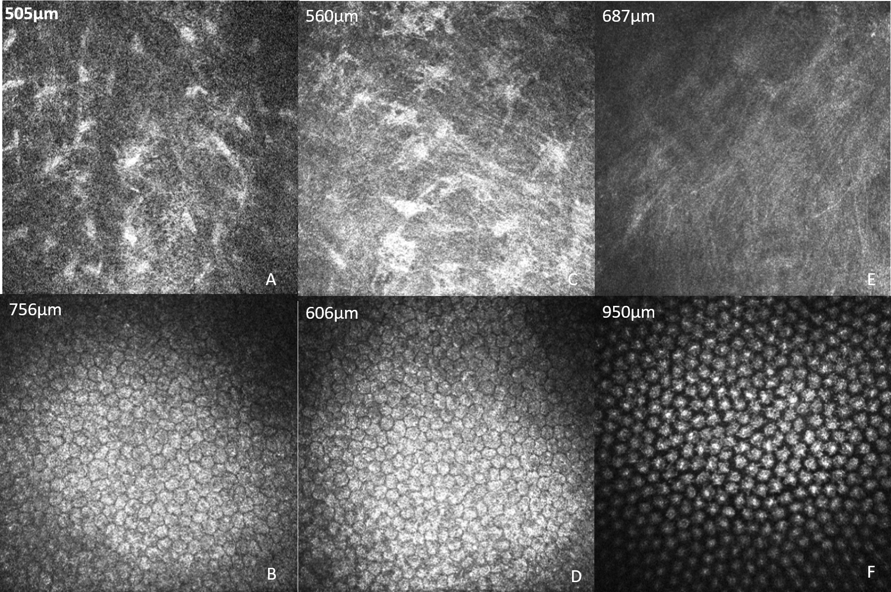
 Figure 2 of
Song, Mol Vis 2017; 23:504-513.
Figure 2 of
Song, Mol Vis 2017; 23:504-513.