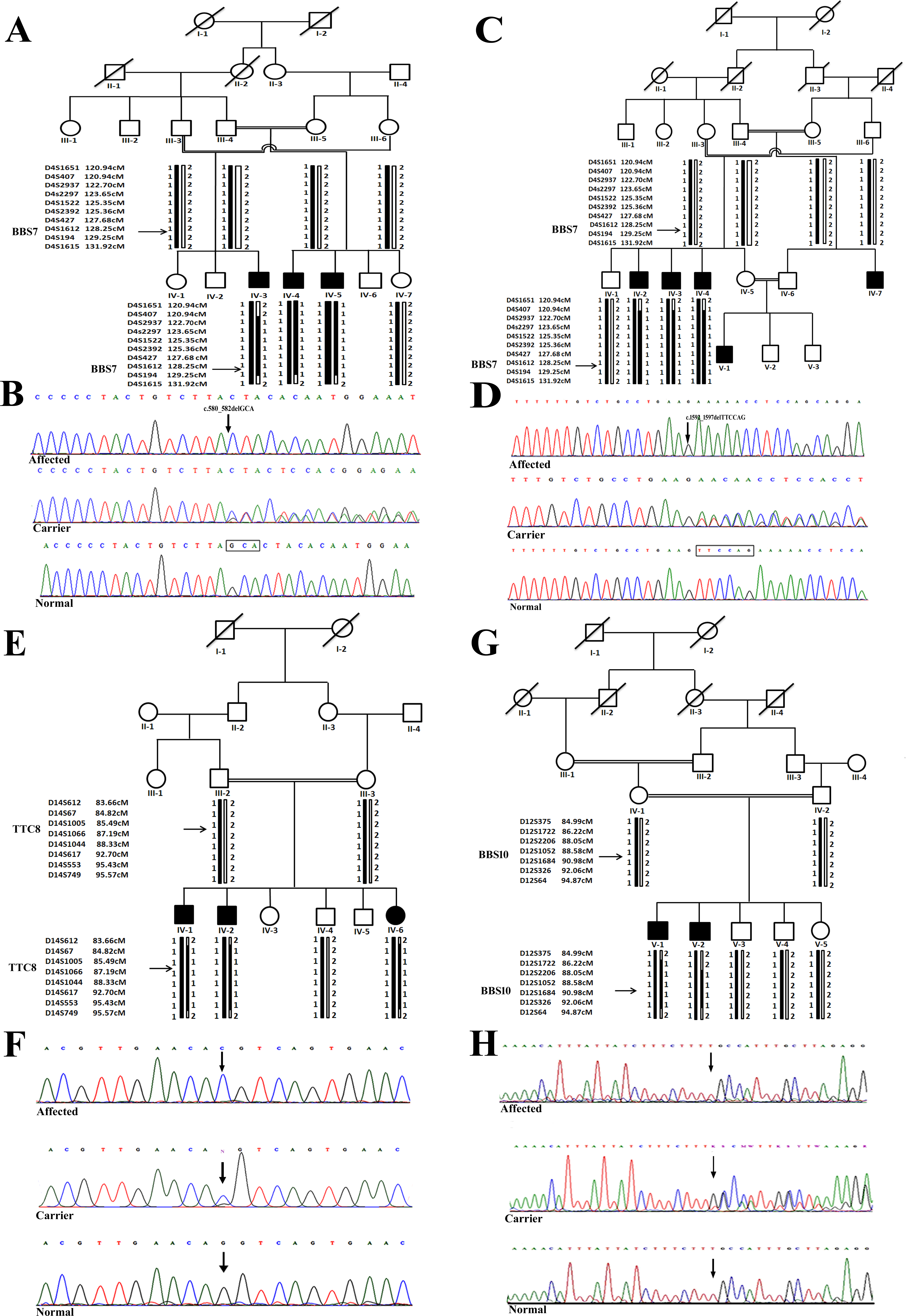
Figure 1. Pedigrees and Sanger sequencing results for four families segregating BBS in an autosomal recessive pattern. A: Pedigree of family A. B: Sequence analysis of the BBS7 gene showing a 3 bp deletion at nucleotide position 580–582 (c.580_582delGCA). C: Pedigree of family B. D: Sequence chromatograms of 6 bp deleted variant (c.1592_1597delTTCCAG) in the BBS7 gene. E: Pedigree of family C. F: Sequence analysis of the variant (c.1347G>C) identified in the gene BBS8 in family C. G: Pedigree of family D. H: Sequence chromatogram of the frameshift mutation (c.271_272insT) found in the BBS10 gene in family D. The genotype of individuals for the mutation identified in the respective family, verified with segregation
analysis, is written below each member tested. The upper panel shows the nucleotide sequence in the homozygous affected member,
the middle panel in the heterozygous carrier, and the lower panel in the homozygous normal member in each sequence chromatogram.

 Figure 1 of
Ullah, Mol Vis 2017; 23:482-494.
  Figure 1 of
Ullah, Mol Vis 2017; 23:482-494.