Figure 4. Physiologic neuronal apoptosis occurred in an age-dependent manner during the postnatal development of the rat retina. Neuronal
apoptosis of rat retinas was detected with cleaved caspase-3 immunolabeling (cleaved caspase-3-positive) and with the terminal
deoxynucleotidyl transferase dUTP nick end labeling (TUNEL) assay (TUNEL-positive; n = 5 retinas per group). A: Representative photomicrograph of caspase-3-positive cells (brown) in the rat retinal GCL. Scale bar = 50 μm. B: Respective photomicrograph of TUNEL-positive cells (red) in the rat retinal GCL. Scale bar = 25 μm. C: The mean percentages of caspase-3 positive cells at P3, P7, and P12. D: The percentages of TUNEL-positive cells in the retinal GCL. #p<0.05, ##p<0.01 versus P3, #p<0.05, ##p<0.01 versus P12. One-way
ANOVA followed by Bonferroni’s post-hoc test. ONL, outer nuclear layer; INL, inner nuclear layer; GCL, ganglion cell layer.
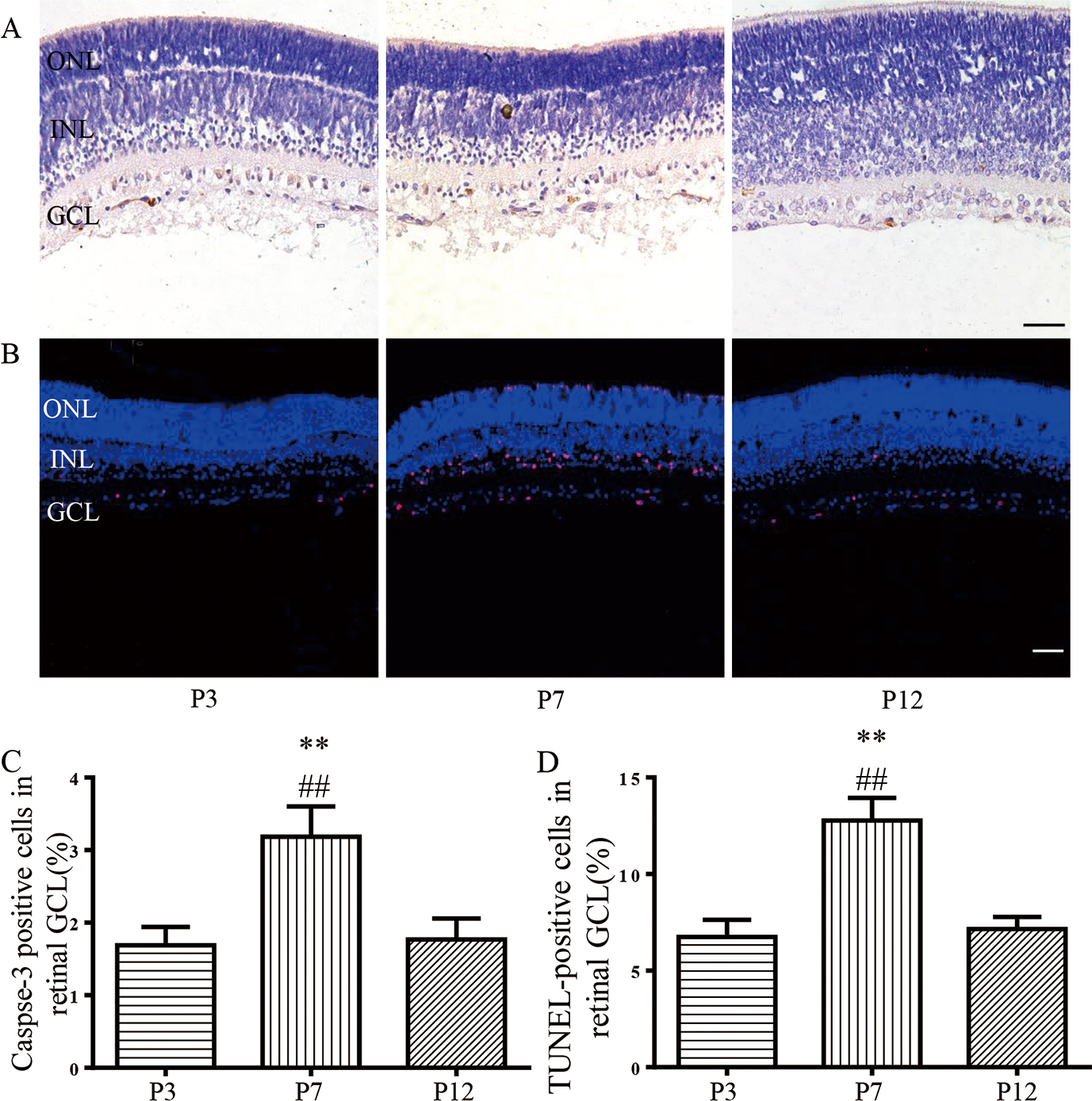
 Figure 4 of
Han, Mol Vis 2017; 23:457-469.
Figure 4 of
Han, Mol Vis 2017; 23:457-469.