Figure 6. Micropatterning of ARPE-19 and hRPE cells. A–C: Scanning electron microscope (SEM) images of through holes of different sizes (A: 100, B: 200, and C: 300 μm in diameter) in the polydimethylsiloxane (PDMS) stencils. The lateral spacing between the holes was 1 mm for all
pattern sizes. D–F: ARPE-19 cells attached to glass coverslips through PDMS stencil holes after 24 h. G–I: hRPE cells on glass coverslips, after the PDMS stencils were removed. Scale bars, A–C = 500 μm; D–I = 200 μm.
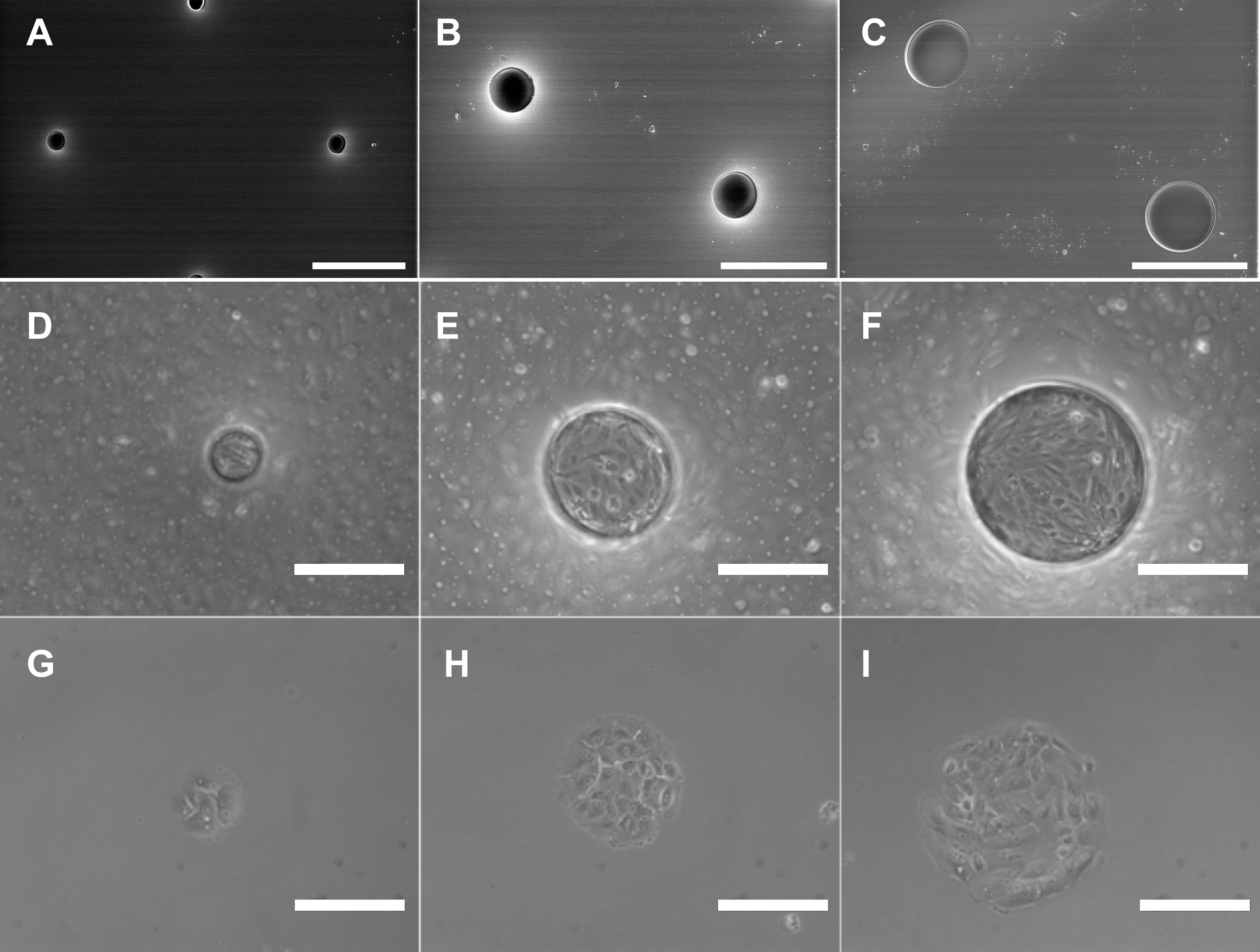
 Figure 6 of
Farjood, Mol Vis 2017; 23:431-446.
Figure 6 of
Farjood, Mol Vis 2017; 23:431-446.