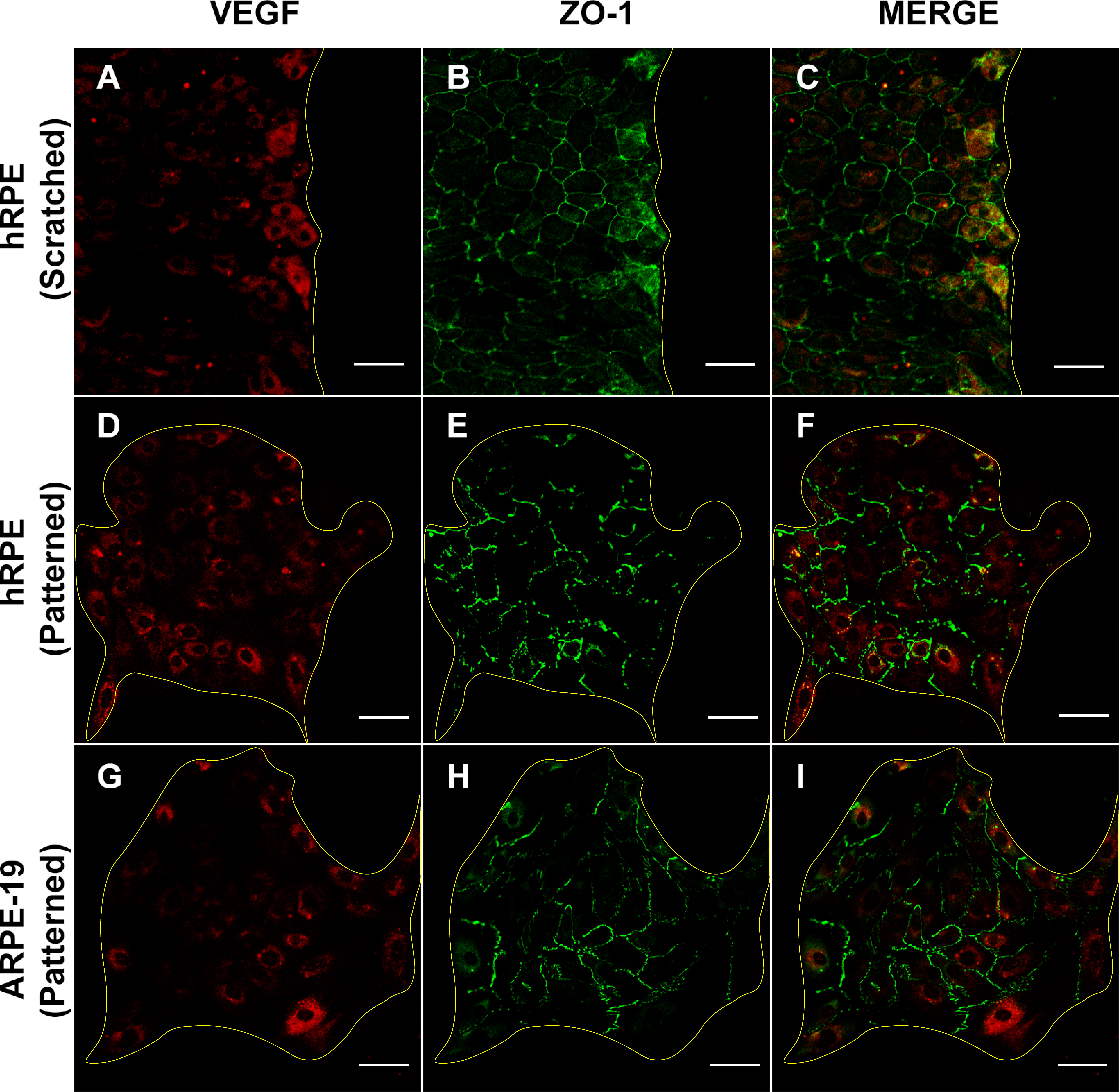
Figure 5. Confocal ICC images of scratched and micropatterned RPE cultures. ZO-1 and VEGF were immunostained in a long-term hRPE culture
(4 weeks after confluency) 24 h after scratching (A-C) and micropatterned hRPE (D-F) and ARPE-19 cells (G-I) 24 hours after removing PDMS stencils. VEGF expression increased in cells proximal to the scratched area (A) and along the periphery of the micropatterns (D, G). This increase in VEGF expression correlated with dislocalization of ZO-1 from intercellular zones to the cytoplasm in both
scratched and micropatterned samples (B, E, H). Red = VEGF; green = ZO-1. Yellow lines indicate the scratch edge (A-C) and micropattern edges (D-I). Scale bar = 50 μm.

 Figure 5 of
Farjood, Mol Vis 2017; 23:431-446.
  Figure 5 of
Farjood, Mol Vis 2017; 23:431-446.