Figure 3. ICC results for confluent ARPE-19 cultures. Cells were immunostained for Claudin-1 (A, B), N-cadherin (C, D), β-catenin (E, F) and ZO-1 (G, H). I-L: Percentage of intercellular junctions covered by the corresponding protein in cultures of ARPE-19 cells grown for 1 day
or 4 weeks after reaching confluency. All junctional proteins, except for ZO-1, had limited localization after 1 day at confluence.
In all cases, junctional localization increased significantly after 4 weeks. Data represent the mean ± standard deviation
for three replicates from three representative confocal images per each time point for each junctional protein (n = 3). *
p<0.05, ** p<0.01. M: Z-stack scan for a long-term (4 weeks after confluency) of ARPE-19 cells grown on porous cell culture inserts. ZO-1 was
localized to the apical junctional areas, while VEGF failed to polarize. Scale bar = 25 μm.
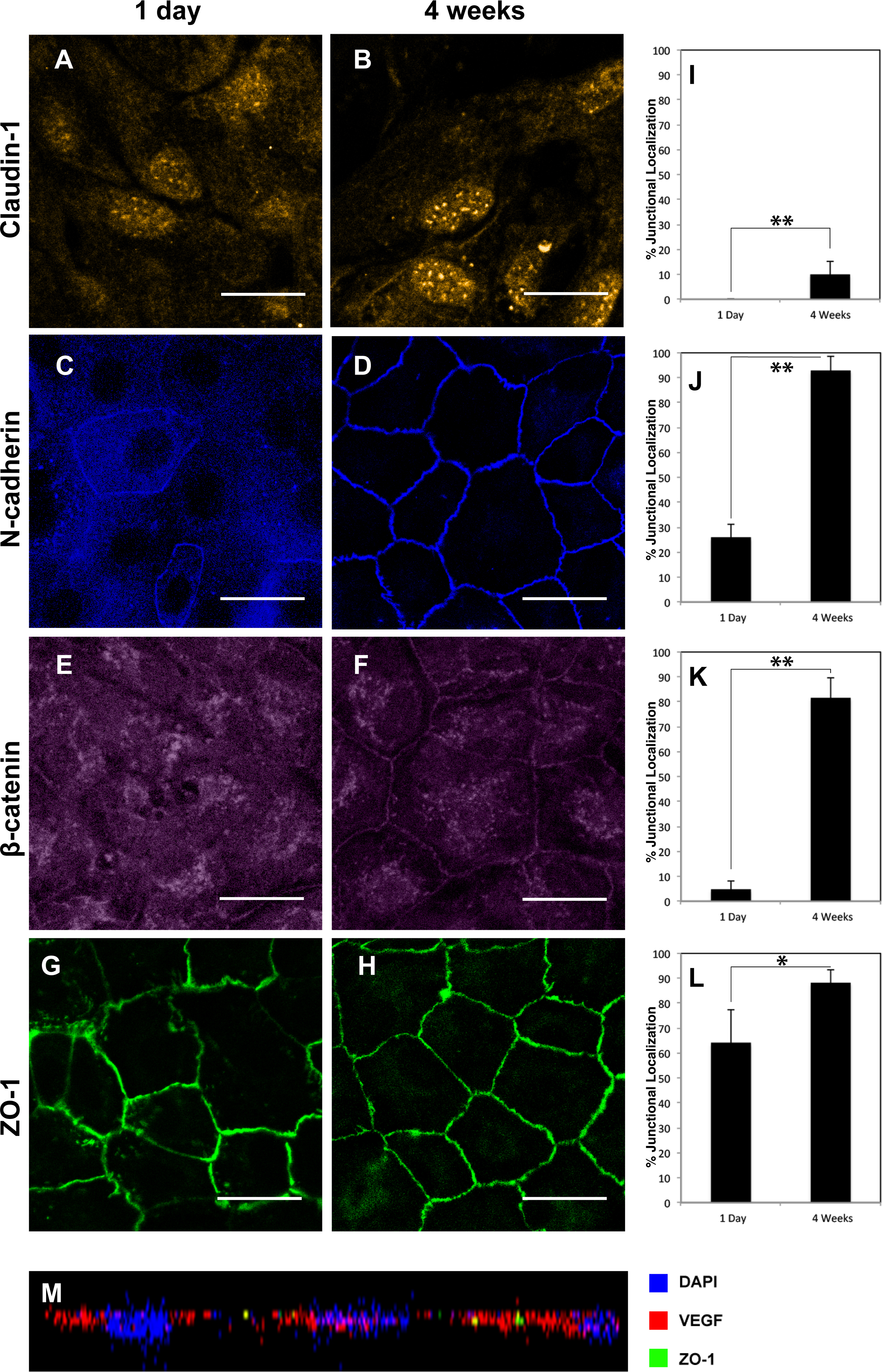
 Figure 3 of
Farjood, Mol Vis 2017; 23:431-446.
Figure 3 of
Farjood, Mol Vis 2017; 23:431-446.