Figure 3. Staining of the separations of porcine vitreous inoculated with Staphylococcus aureus over 12 h. After homogenizing and centrifuging, Gram staining (A, B) and Coomassie Blue staining (C, D) were conducted to observe the vitreous supernatants (A, C) and pellets (B, D). In A and B, the scale bar is equal to 10 µm, and in C and D, the scale bar is equal to 20 µm.
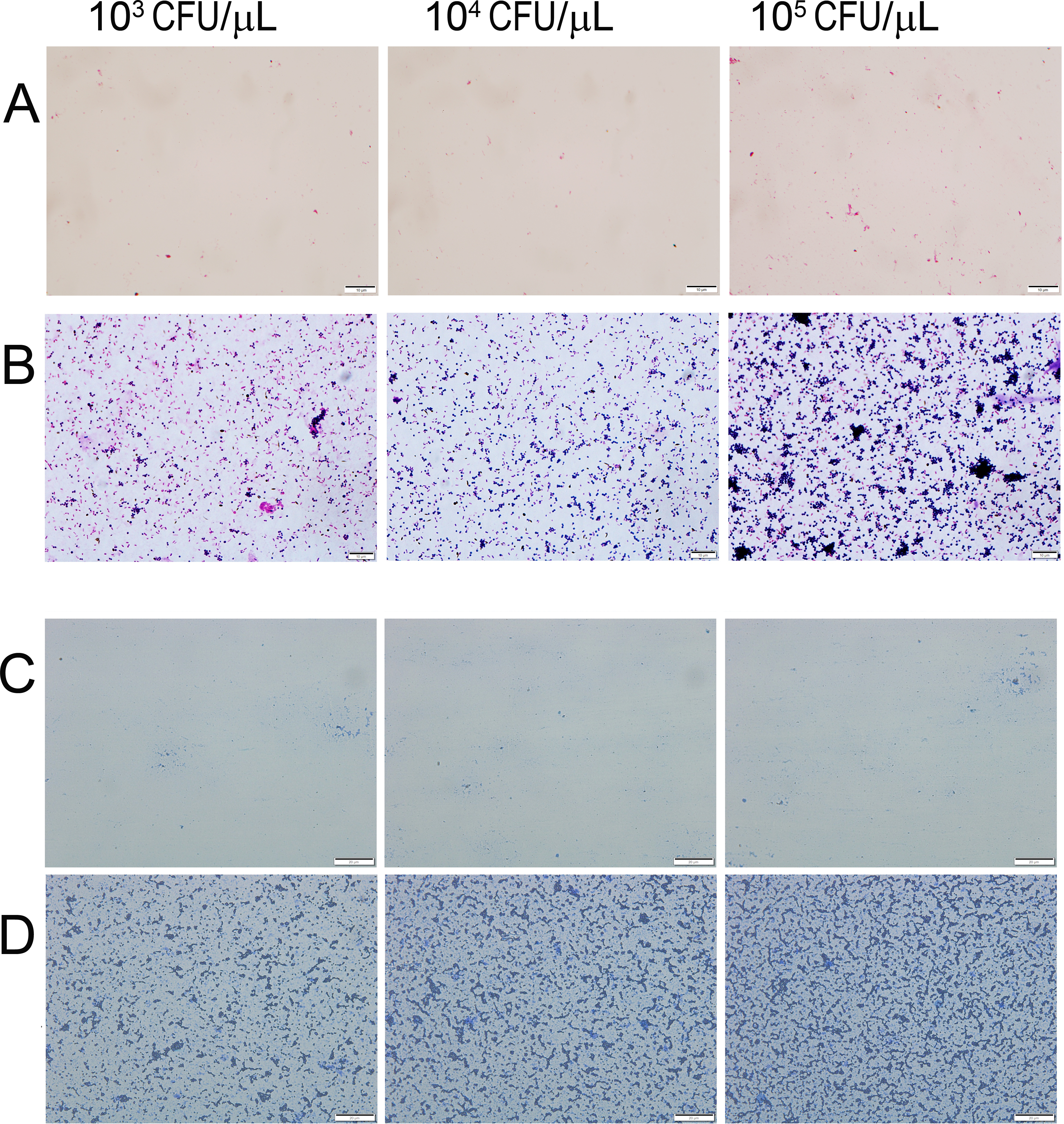
 Figure 3 of
Song, Mol Vis 2017; 23:407-415.
Figure 3 of
Song, Mol Vis 2017; 23:407-415.