Figure 2. Histological evaluation of the eyes from the mice treated with or without LPS. Representative images (A, C) show infiltrating inflammatory cells in the posterior chamber of the eyes in the control group at the 24th hour. Representative
images (B, D) show infiltrating inflammatory cells in the posterior chamber of the eyes in the lipopolysaccharide (LPS) group at the 24th
hour after LPS injection. Magnification = 10X for A and B and 200X for C and D (n = 3).
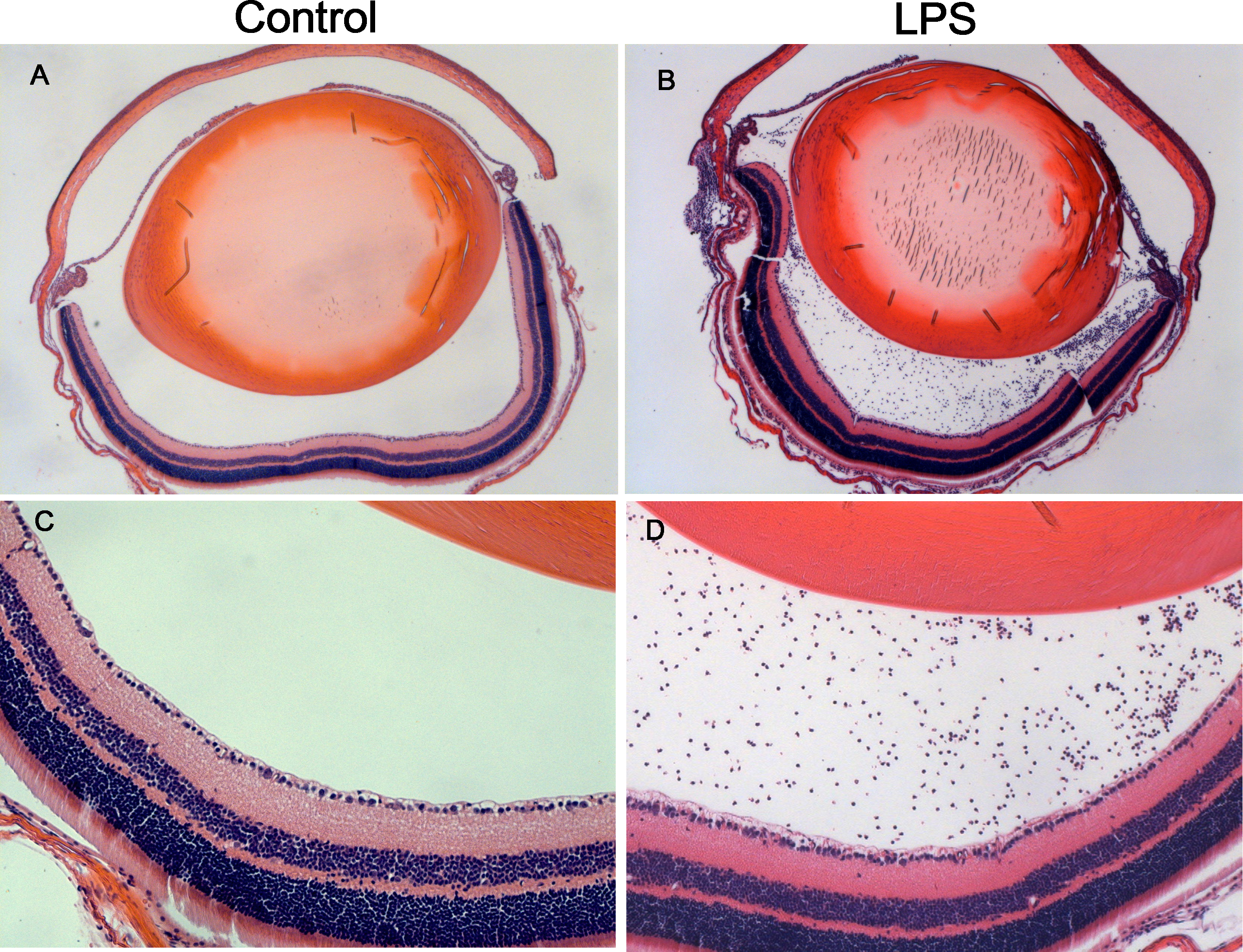
 Figure 2 of
Qiu, Mol Vis 2017; 23:395-406.
Figure 2 of
Qiu, Mol Vis 2017; 23:395-406.