Figure 6. Protective effects of PN-1 and PEDF proteins and peptides in cultured cells. R28 cells in serum-free media were treated with
serpin proteins and peptides, as indicated, for 48 h. Cytoprotection was assayed by counting the terminal deoxynucleotidyl
transferase (TdT) dUTP nick-end labeling (TUNEL)-positive nuclei (green). Cells were fixed and processed for TUNEL staining
and counterstained with Hoechst dye for the nucleus. A: Representative images from each condition show the TUNEL-positive nuclei (green) and the Hoechst-stained nuclei (blue, total
cells). Concentrations of proteins and peptides were 2.5 nM. Bar = 20 µm. B: Quantification of the protective effects by PN-1 proteins treated as in A. Each bar corresponds to the average of the percentage of TUNEL-positive nuclei per the total number of cells normalized
to the cells treated with serum-free medium (None) as 100%. Shown are data from three independent experiments each performed
with duplicate wells ± standard error of the mean (SEM; error bars). C: Cells were pretreated with atglistatin (3.5 µM) for 1 h before a 48-h treatment with or without indicated concentrations
of PEDF, PN-1, and PN-1-17mer. Quantification of TUNEL-positive nuclei is shown. Each bar corresponds to the average of the
percentage of the TUNEL-positive nuclei per the total number of cells from two independent batches of cells and each performed
with duplicate wells per assay ± standard deviation (SD) **, p<0.005; ***, p<0.0005; NS, not significant.
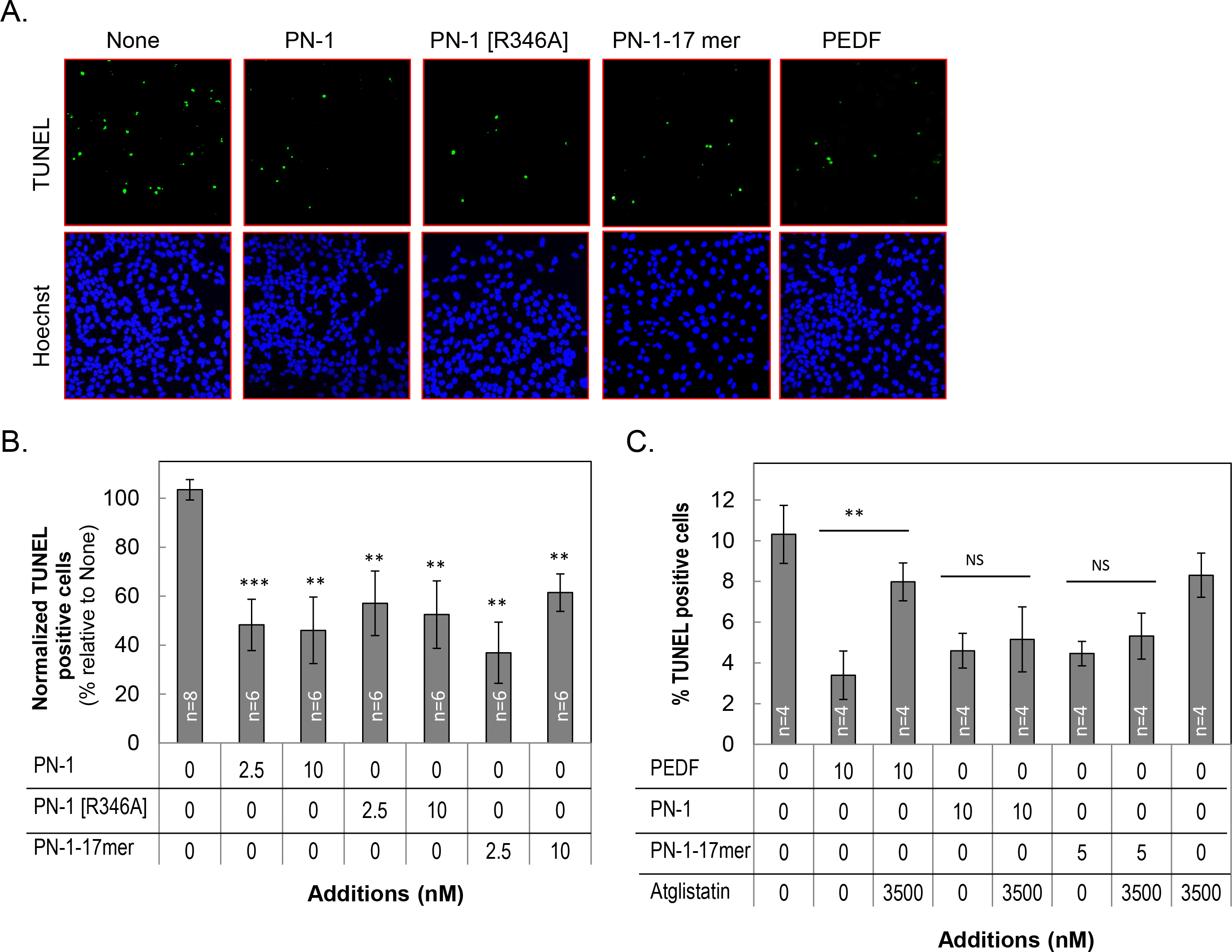
 Figure 6 of
Winokur, Mol Vis 2017; 23:372-384.
Figure 6 of
Winokur, Mol Vis 2017; 23:372-384.