Figure 4. PN-1 protein in human ARPE-19 cell cultures. Protein samples were resolved in a polyacrylamide gel, transferred to nitrocellulose
membranes, and immunostained with antibodies. ARPE-19 cells were cultured in serum-free media for 5 days. Conditioned media
were removed, and a wash with PBS containing 2 M NaCl was performed. Cell lysates were prepared from the cells. Photos of
blots with samples loaded in two gels in duplicate as follows: ARPE-19 conditioned media from serum-free media (lane 1), 2
M NaCl wash (lane 2), bacterially derived recombinant human PN-1 (rhuPN-1, lane 3 top), mammalian-derived, recombinant human
PEDF (rhuPEDF, lane 3 bottom), and ARPE-19 cell lysate (lane 4). The top blot was immunostained with Ab-PN-1 and the bottom
with Ab-PEDF. For both panels, the migration positions of the MW markers are shown to the left. The migration positions for
rhuPN-1 and rhuPEDF are shown to the right. The asterisk points to the migration position of mature glycosylated PN-1.
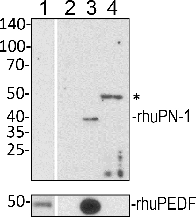
 Figure 4 of
Winokur, Mol Vis 2017; 23:372-384.
Figure 4 of
Winokur, Mol Vis 2017; 23:372-384.