Figure 1. Isolectin GS-IB4 and FITC-dextran staining shows the distribution of healthy and neovascular blood vessels in the retina.
Retinal whole mounts stained with isolectin GS-IB4 and FITC-dextran in eyes from healthy mice and mice treated with hyperoxia.
Representative images with isolectin GS-IB4 (A, C, E, G, I) and FITC (B, D, F, H, J). A, B: Staining in eyes from healthy mice. C, D: Staining in eyes from mice treated with hyperoxia. E, F: Magnified images showing the distribution of the retinal blood vessels. G, H: Magnified images showing neovascular tufts. I: Image produced in Photoshop. The neovascular areas are green, and the obliterative area is yellow. J: Image produced in Photoshop. The neovascular areas are red, and the obliterative area is yellow.
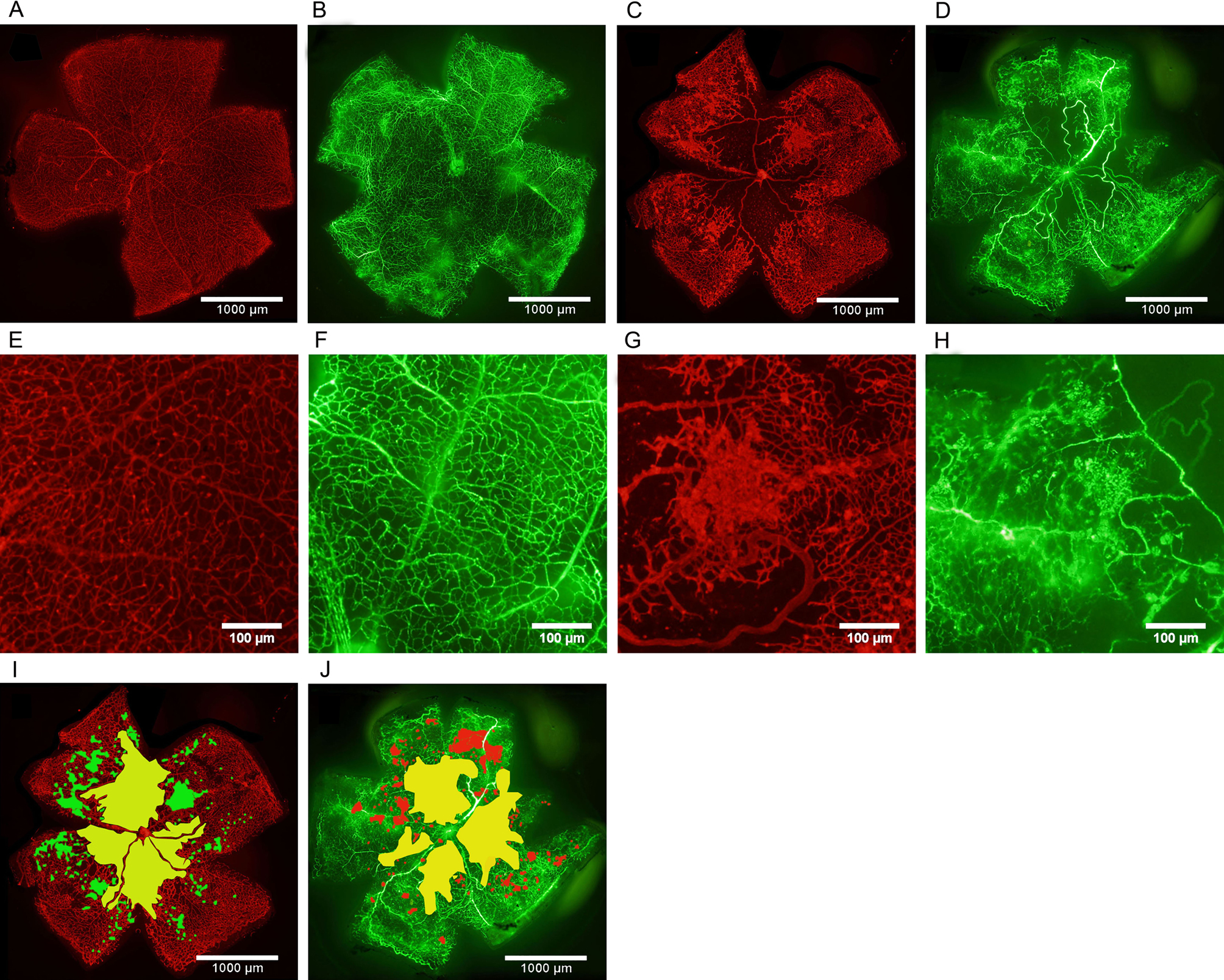
 Figure 1 of
Zhang, Mol Vis 2017; 23:346-355.
Figure 1 of
Zhang, Mol Vis 2017; 23:346-355.